


Crystal structure of glycerol dehydrogenase from Schyzosaccharomyces pombe
Mulichak, A.M.To be published.
Experimental Data Snapshot
wwPDB Validation 3D Report Full Report

Entity ID: 1 | |||||
|---|---|---|---|---|---|
| Molecule | Chains | Sequence Length | Organism | Details | Image |
| glycerol dehydrogenase | 450 | Schizosaccharomyces pombe | Mutation(s): 0 EC: 1.1.1.6 | 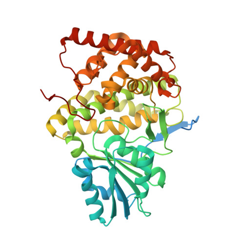 | |
UniProt | |||||
Entity Groups | |||||
| Sequence Clusters | 30% Identity50% Identity70% Identity90% Identity95% Identity100% Identity | ||||
| UniProt Group | O13702 | ||||
Sequence AnnotationsExpand | |||||
Reference Sequence | |||||
| Ligands 4 Unique | |||||
|---|---|---|---|---|---|
| ID | Chains | Name / Formula / InChI Key | 2D Diagram | 3D Interactions | |
| GOL Download:Ideal Coordinates CCD File | G [auth A], H [auth A], K [auth B] | GLYCEROL C3 H8 O3 PEDCQBHIVMGVHV-UHFFFAOYSA-N |  | ||
| ALA Download:Ideal Coordinates CCD File | E [auth A], F [auth A] | ALANINE C3 H7 N O2 QNAYBMKLOCPYGJ-REOHCLBHSA-N |  | ||
| ZN Download:Ideal Coordinates CCD File | C [auth A], I [auth B] | ZINC ION Zn PTFCDOFLOPIGGS-UHFFFAOYSA-N |  | ||
| K Download:Ideal Coordinates CCD File | D [auth A], J [auth B] | POTASSIUM ION K NPYPAHLBTDXSSS-UHFFFAOYSA-N |  | ||
| Length ( Å ) | Angle ( ˚ ) |
|---|---|
| a = 105.98 | α = 90 |
| b = 105.98 | β = 90 |
| c = 140.63 | γ = 90 |
| Software Name | Purpose |
|---|---|
| MAR345 | data collection |
| SCALEPACK | data scaling |
| SOLVE | phasing |
| CNS | refinement |


