1T6U

Nickel Superoxide Dismutase (NiSOD) Native 1.30 A Structure


Experimental Data Snapshot

  • Method: X-RAY DIFFRACTION
  • Resolution: 1.30 Å
  • R-Value Free: 0.201 
  • R-Value Work: 0.158 

wwPDB Validation   3D Report Full Report


This is version 1.4 of the entry. See complete history


Literature

Nickel superoxide dismutase structure and mechanism.

Barondeau, D.P.Kassmann, C.J.Bruns, C.K.Tainer, J.A.Getzoff, E.D.

(2004) Biochemistry 43: 8038-8047

  • DOI: https://doi.org/10.1021/bi0496081
  • Primary Citation of Related Structures:  
    1T6I, 1T6Q, 1T6U

  • PubMed Abstract: 

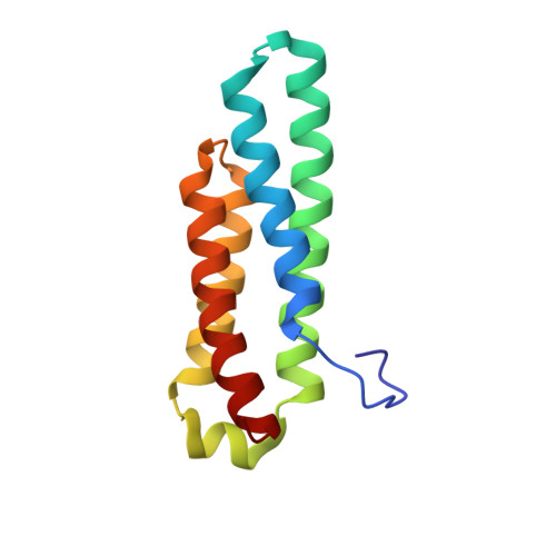
    The 1.30 A resolution crystal structure of nickel superoxide dismutase (NiSOD) identifies a novel SOD fold, assembly, and Ni active site. NiSOD is a hexameric assembly of right-handed 4-helix bundles of up-down-up-down topology with N-terminal hooks chelating the active site Ni ions. This newly identified nine-residue Ni-hook structural motif (His-Cys-X-X-Pro-Cys-Gly-X-Tyr) provides almost all interactions critical for metal binding and catalysis, and thus will likely be diagnostic of NiSODs. Conserved lysine residues are positioned for electrostatic guidance of the superoxide anion to the narrow active site channel. Apo structures show that the Ni-hook motif is unfolded prior to metal binding. The active site Ni geometry cycles from square planar Ni(II), with thiolate (Cys2 and Cys6) and backbone nitrogen (His1 and Cys2) ligands, to square pyramidal Ni(III) with an added axial His1 side chain ligand, consistent with electron paramagentic resonance spectroscopy. Analyses of the three NiSOD structures and comparisons to the Cu,Zn and Mn/Fe SODs support specific molecular mechanisms for NiSOD maturation and catalysis, and identify important structure-function relationships conserved among SODs.


  • Organizational Affiliation

    Department of Molecular Biology, The Skaggs Institute for Chemical Biology, The Scripps Research Institute, 10550 North Torrey Pines Road, La Jolla, California 92037, USA.


Macromolecules
Find similar proteins by:  (by identity cutoff)  |  3D Structure
Entity ID: 1
MoleculeChains Sequence LengthOrganismDetailsImage
Superoxide dismutase [Ni]
A, B, C, D, E
A, B, C, D, E, F, G, H, I, J, K, L
117Streptomyces coelicolorMutation(s): 1 
Gene Names: SODNSOD1SCO52542SC7G11.16C
EC: 1.15.1.1
UniProt
Find proteins for P80735 (Streptomyces coelicolor (strain ATCC BAA-471 / A3(2) / M145))
Explore P80735 
Go to UniProtKB:  P80735
Entity Groups  
Sequence Clusters30% Identity50% Identity70% Identity90% Identity95% Identity100% Identity
UniProt GroupP80735
Sequence Annotations
Expand
  • Reference Sequence
Small Molecules
Ligands 1 Unique
IDChains Name / Formula / InChI Key2D Diagram3D Interactions
NI
Query on NI

Download Ideal Coordinates CCD File 
M [auth A]
N [auth B]
O [auth C]
P [auth D]
Q [auth E]
M [auth A],
N [auth B],
O [auth C],
P [auth D],
Q [auth E],
R [auth F],
S [auth G],
T [auth H],
U [auth I],
V [auth J],
W [auth K],
X [auth L]
NICKEL (II) ION
Ni
VEQPNABPJHWNSG-UHFFFAOYSA-N
Experimental Data & Validation

Experimental Data

  • Method: X-RAY DIFFRACTION
  • Resolution: 1.30 Å
  • R-Value Free: 0.201 
  • R-Value Work: 0.158 
  • Space Group: P 1 21 1
Unit Cell:
Length ( Å )Angle ( ˚ )
a = 64.147α = 90
b = 111.68β = 94.51
c = 106.834γ = 90
Software Package:
Software NamePurpose
SHELXmodel building
SHELXL-97refinement
DENZOdata reduction
SCALEPACKdata scaling
AMoREphasing

Structure Validation

View Full Validation Report


Entry History 

Deposition Data

Revision History  (Full details and data files)

  • Version 1.0: 2004-07-13
    Type: Initial release
  • Version 1.1: 2008-04-30
    Changes: Version format compliance
  • Version 1.2: 2011-07-13
    Changes: Version format compliance
  • Version 1.3: 2021-10-27
    Changes: Database references, Derived calculations
  • Version 1.4: 2023-08-23
    Changes: Data collection, Refinement description