Inhibitor coordination interactions in the binuclear manganese cluster of arginase
Cama, E., Pethe, S., Boucher, J.-L., Han, S., Emig, F.A., Ash, D.E., Viola, R.E., Mansuy, D., Christianson, D.W.(2004) Biochemistry 43: 8987-8999
- PubMed: 15248756 Search on PubMed
- DOI: https://doi.org/10.1021/bi0491705
- Primary Citation Related Structures:
1T4P, 1T4R, 1T4S, 1T4T, 1T5G - PubMed Abstract:
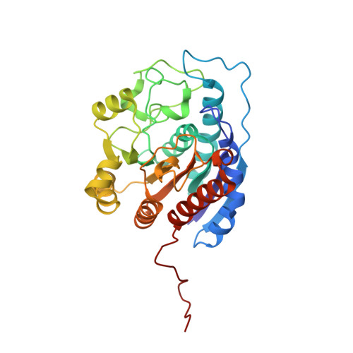
Arginase is a manganese metalloenzyme that catalyzes the hydrolysis of L-arginine to form L-ornithine and urea. The structure and stability of the binuclear manganese cluster are critical for catalytic activity as it activates the catalytic nucleophile, metal-bridging hydroxide ion, and stabilizes the tetrahedral intermediate and its flanking states. Here, we report X-ray structures of a series of inhibitors bound to the active site of arginase, and each inhibitor exploits a different mode of coordination with the Mn(2+)(2) cluster. Specifically, we have studied the binding of fluoride ion (F(-); an uncompetitive inhibitor) and L-arginine, L-valine, dinor-N(omega)-hydroxy-L-arginine, descarboxy-nor-N(omega)-hydroxy-L-arginine, and dehydro-2(S)-amino-6-boronohexanoic acid. Some inhibitors, such as fluoride ion, dinor-N(omega)-hydroxy-L-arginine, and dehydro-2(S)-amino-6-boronohexanoic acid, cause the net addition of one ligand to the Mn(2+)(2) cluster. Other inhibitors, such as descarboxy-nor-N(omega)-hydroxy-L-arginine, simply displace the metal-bridging hydroxide ion of the native enzyme and do not cause any net change in the metal coordination polyhedra. The highest affinity inhibitors displace the metal-bridging hydroxide ion (and sometimes occupy a Mn(2+)(A) site found vacant in the native enzyme) and maintain a conserved array of hydrogen bonds with their alpha-amino and -carboxylate groups.
- Roy and Diana Vagelos Laboratories, Department of Chemistry, University of Pennsylvania, Philadelphia, Pennsylvania 19104-6323, USA.
Organizational Affiliation: