Solution Structure of AF-6 PDZ Domain and Its Interaction with the C-terminal Peptides from Neurexin and Bcr
Zhou, H., Xu, Y., Yang, Y., Huang, A., Wu, J., Shi, Y.(2005) J Biological Chem 280: 13841-13847
- PubMed: 15684424 Search on PubMed
- DOI: https://doi.org/10.1074/jbc.M411065200
- Primary Citation Related Structures:
1T2M - PubMed Abstract:
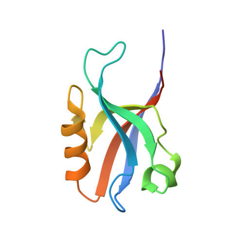
AF-6 is a key molecule essential for structure organization of cell-cell junction of polarized epithelia. It belongs to a novel cell-cell adhesion system. The AF-6 PDZ domain mediates interactions by binding to a specific amino acid sequence in target proteins. Here we report the solution structure of the AF-6 PDZ domain determined by NMR. Previously, the AF-6 PDZ domain was considered to be a class II PDZ domain. However we found that a unique hydrophilic amino acid, Gln70, at position alphaB1 makes the alphaB/betaB groove of the AF-6 PDZ domain significantly different from that of the canonical class II PDZ domain. The AF-6 PDZ domain does not have the second hydrophobic binding pocket, and the N-terminal end of alphaB is closer to betaB. Using BIACORE and NMR chemical shift perturbation experiments, we have studied the binding characteristics of the PDZ domain to the C-terminal peptide of Neurexin, KKNKDKEYYV, and that of Bcr, KRQSILFSTEV. The C-terminal peptide of Neurexin is a class II ligand, whereas that of Bcr is a class I ligand. The dissociation constants of these ligands were 4.08 x 10(-7) and 2.23 x 10(-6) m, respectively. Each of the four C-terminal positions in Neurexin and Bcr may contribute to the interaction. The three-dimensional models of the AF-6 PDZ-Neurexin C-terminal peptide complex and the AF-6 PDZ-Bcr C-terminal peptide complex were built up by molecular dynamics simulations. Unlike the canonical class II PDZ domain, Ala74 at alphaB5 rather than the residue at alphaB1 makes direct hydrophobic contact with the side chain of Tyr at the -2 position of the ligand.
- Hefei National Laboratory for Physical Sciences at Microscale, School of Life Science, University of Science and Technology of China, Hefei, Anhui 230026, People's Republic of China.
Organizational Affiliation: