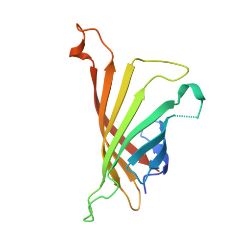
Structural studies of the streptavidin binding loop.
Freitag, S., Le Trong, I., Klumb, L., Stayton, P.S., Stenkamp, R.E.(1997) Protein Sci 6: 1157-1166
- PubMed: 9194176 Search on PubMedSearch on PubMed Central
- DOI: https://doi.org/10.1002/pro.5560060604
- Primary Citation Related Structures:
1SWA, 1SWB, 1SWC, 1SWD, 1SWE - PubMed Abstract:
The streptavidin-biotin complex provides the basis for many important biotechnological applications and is an interesting model system for studying high-affinity protein-ligand interactions. We report here crystallographic studies elucidating the conformation of the flexible binding loop of streptavidin (residues 45 to 52) in the unbound and bound forms. The crystal structures of unbound streptavidin have been determined in two monoclinic crystal forms. The binding loop generally adopts an open conformation in the unbound species. In one subunit of one crystal form, the flexible loop adopts the closed conformation and an analysis of packing interactions suggests that protein-protein contacts stabilize the closed loop conformation. In the other crystal form all loops adopt an open conformation. Co-crystallization of streptavidin and biotin resulted in two additional, different crystal forms, with ligand bound in all four binding sites of the first crystal form and biotin bound in only two subunits in a second. The major change associated with binding of biotin is the closure of the surface loop incorporating residues 45 to 52. Residues 49 to 52 display a 3(10) helical conformation in unbound subunits of our structures as opposed to the disordered loops observed in other structure determinations of streptavidin. In addition, the open conformation is stabilized by a beta-sheet hydrogen bond between residues 45 and 52, which cannot occur in the closed conformation. The 3(10) helix is observed in nearly all unbound subunits of both the co-crystallized and ligand-free structures. An analysis of the temperature factors of the binding loop regions suggests that the mobility of the closed loops in the complexed structures is lower than in the open loops of the ligand-free structures. The two biotin bound subunits in the tetramer found in the MONO-b1 crystal form are those that contribute Trp 120 across their respective binding pockets, suggesting a structural link between these binding sites in the tetramer. However, there are no obvious signatures of binding site communication observed upon ligand binding, such as quaternary structure changes or shifts in the region of Trp 120. These studies demonstrate that while crystallographic packing interactions can stabilize both the open and closed forms of the flexible loop, in their absence the loop is open in the unbound state and closed in the presence of biotin. If present in solution, the helical structure in the open loop conformation could moderate the entropic penalty associated with biotin binding by contributing an order-to-disorder component to the loop closure.
- Department of Biological Structure, University of Washington, Seattle 98195-7742, USA.
Organizational Affiliation: