1SVZ

Crystal structure of the single-chain Fv fragment 1696 in complex with the epitope peptide corresponding to N-terminus of HIV-2 protease


Experimental Data Snapshot

  • Method: X-RAY DIFFRACTION
  • Resolution: 1.89 Å
  • R-Value Free: 0.239 
  • R-Value Work: 0.196 
  • R-Value Observed: 0.196 

wwPDB Validation   3D Report Full Report


This is version 1.3 of the entry. See complete history


Literature

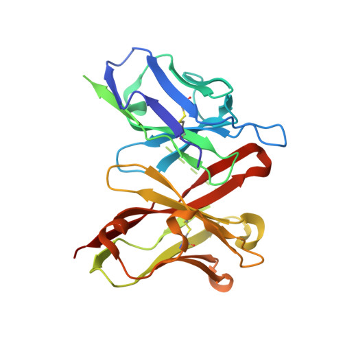
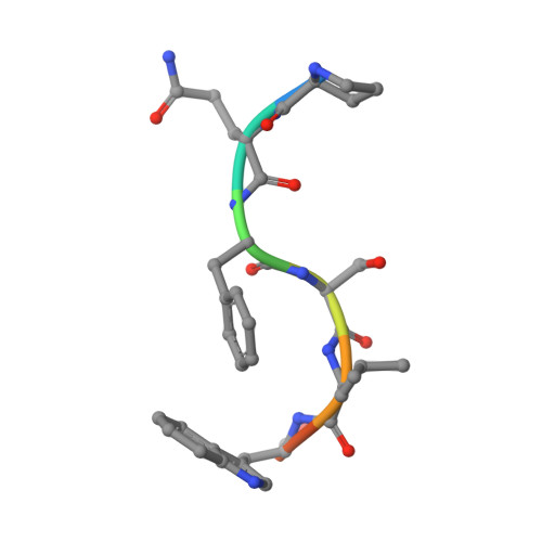
Crystal structure of a cross-reaction complex between an anti-HIV-1 protease antibody and an HIV-2 protease peptide

Rezacova, P.Brynda, J.Lescar, J.Fabry, M.Horejsi, M.Sieglova, I.Sedlacek, J.Bentley, G.A.

(2005) J Struct Biol 149: 332-337

  • DOI: https://doi.org/10.1016/j.jsb.2004.11.009
  • Primary Citation of Related Structures:  
    1SVZ

  • PubMed Abstract: 
  • The monoclonal antibody 1696, elicited by HIV-1 protease, inhibits the activity of both HIV-1 and HIV-2 proteases with inhibition constants in the low nanomolar range. The antibody cross-reacts with peptides derived from the N-terminal region of both proteases ...

    The monoclonal antibody 1696, elicited by HIV-1 protease, inhibits the activity of both HIV-1 and HIV-2 proteases with inhibition constants in the low nanomolar range. The antibody cross-reacts with peptides derived from the N-terminal region of both proteases. The crystal structure of the recombinant single-chain Fv fragment of 1696 complexed with an N-terminal peptide from the HIV-2 protease has been determined at 1.88A resolution. Interactions of the peptide with scFv1696 are compared with the previously reported structure of scFv1696 in complex with the corresponding peptide from HIV-1 protease. The origin of cross-reactivity of mAb1696 with HIV proteases is discussed.


    Organizational Affiliation

    Department of Recombinant Expression and Structural Biology, Institute of Molecular Genetics, Academy of Sciences of the Czech Republic, Flemingovo nam. 2, 166 37 Prague 6, Czech Republic. rezacova@img.cas.nz


Macromolecules
Find similar proteins by:  (by identity cutoff)  |  3D Structure
Entity ID: 1
MoleculeChainsSequence LengthOrganismDetailsImage
single-chain Fv fragment 1696
A, B
247Mus musculusMutation(s): 0 
UniProt
Find proteins for P01631 (Mus musculus)
Explore P01631 
Go to UniProtKB:  P01631
Entity Groups  
Sequence Clusters30% Identity50% Identity70% Identity90% Identity95% Identity100% Identity
UniProt GroupP01631
Protein Feature View
Expand
  • Reference Sequence

Find similar proteins by:  Sequence   |   3D Structure  

Entity ID: 2
MoleculeChainsSequence LengthOrganismDetailsImage
epitope peptide corresponding to N-terminus of HIV-2 protease
C, D
8N/AMutation(s): 0 
Protein Feature View
Expand
  • Reference Sequence
Experimental Data & Validation

Experimental Data

  • Method: X-RAY DIFFRACTION
  • Resolution: 1.89 Å
  • R-Value Free: 0.239 
  • R-Value Work: 0.196 
  • R-Value Observed: 0.196 
  • Space Group: P 1 21 1
Unit Cell:
Length ( Å )Angle ( ˚ )
a = 44.184α = 90
b = 56.413β = 102.13
c = 88.304γ = 90
Software Package:
Software NamePurpose
DENZOdata reduction
SCALEPACKdata scaling
AMoREphasing
CNSrefinement

Structure Validation

View Full Validation Report


Entry History 

Deposition Data

Revision History  (Full details and data files)

  • Version 1.0: 2005-03-01
    Type: Initial release
  • Version 1.1: 2008-04-30
    Changes: Version format compliance
  • Version 1.2: 2011-07-13
    Changes: Version format compliance
  • Version 1.3: 2023-08-23
    Changes: Data collection, Database references, Refinement description