1SUP | pdb_00001sup

SUBTILISIN BPN' AT 1.6 ANGSTROMS RESOLUTION: ANALYSIS OF DISCRETE DISORDER AND COMPARISON OF CRYSTAL FORMS


Experimental Data Snapshot

  • Method: X-RAY DIFFRACTION
  • Resolution: 1.60 Å
  • R-Value Observed: 
    0.168 (Depositor) 

wwPDB Validation 3D Report Full Report

Validation slider image for 1SUP

This is version 1.4 of the entry. See complete history

Literature

Subtilisin BPN' at 1.6 A resolution: analysis for discrete disorder and comparison of crystal forms.

Gallagher, T.Oliver, J.Bott, R.Betzel, C.Gilliland, G.L.

(1996) Acta Crystallogr D Biol Crystallogr 52: 1125-1135

  • DOI: https://doi.org/10.1107/S0907444996007500
  • Primary Citation Related Structures: 
    1SUP

  • PubMed Abstract: 

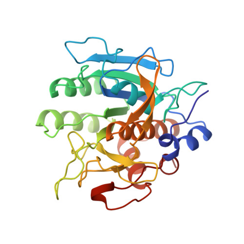
    The three-dimensional structure of the serine protease subtilisin BPN' (SBT) has been refined at 1.6 A resolution in space group C2 to a final R value of 0.17. 17 regions of discrete disorder have been identified and analyzed. Two of these are dual-conformation peptide units; the remainder involve alternate rotamers of side chains either alone or in small clusters. The structure is compared with previously reported high-resolution models of SBT in two other space groups, P2(1)2(1)2(1) and P2(1). Apart from the surface, there are no significant variations in structure among the three crystal forms. Structural variations observed at the protein surface occur predominantly in regions of protein-protein contact. The crystal packing arrangements in the three space groups are compared.


  • Organizational Affiliation
    • Center for Advanced Reseach in Biotechnology, University of Maryland Biotechnology Institute, National Institute of Standards and Technology, Rockville, MD 20850, USA.

Macromolecule Content 

  • Total Structure Weight: 27.79 kDa 
  • Atom Count: 2,224 
  • Modeled Residue Count: 275 
  • Deposited Residue Count: 275 
  • Unique protein chains: 1

Macromolecules

Find similar proteins by:|  3D Structure
Entity ID: 1
MoleculeChains  Sequence LengthOrganismDetailsImage
SUBTILISIN BPN'275Bacillus amyloliquefaciensMutation(s): 0 
EC: 3.4.21.62
UniProt
Find proteins for P00782 (Bacillus amyloliquefaciens)
Explore P00782 
Go to UniProtKB:  P00782
Entity Groups
Sequence Clusters30% Identity50% Identity70% Identity90% Identity95% Identity100% Identity
UniProt GroupP00782
Sequence Annotations
Expand
Reference Sequence

Small Molecules

Ligands 3 Unique
IDChains Name / Formula / InChI Key2D Diagram3D Interactions
PMS

Query on PMS


Download:Ideal Coordinates CCD File
D [auth A]phenylmethanesulfonic acid
C7 H8 O3 S
NIXKBAZVOQAHGC-UHFFFAOYSA-N
CA

Query on CA


Download:Ideal Coordinates CCD File
B [auth A]CALCIUM ION
Ca
BHPQYMZQTOCNFJ-UHFFFAOYSA-N
NA

Query on NA


Download:Ideal Coordinates CCD File
C [auth A]SODIUM ION
Na
FKNQFGJONOIPTF-UHFFFAOYSA-N

Experimental Data & Validation

Experimental Data

  • Method: X-RAY DIFFRACTION
  • Resolution: 1.60 Å
  • R-Value Observed: 0.168 (Depositor) 
Space Group: C 1 2 1
Unit Cell:
Length ( Å )Angle ( ˚ )
a = 66.56α = 90
b = 54.15β = 91.87
c = 62.73γ = 90
Software Package:
Software NamePurpose
PROLSQrefinement

Structure Validation

View Full Validation Report


Entry History 

Deposition Data

Revision History  (Full details and data files)

  • Version 1.0: 1995-11-14
    Type: Initial release
  • Version 1.1: 2008-03-03
    Changes: Version format compliance
  • Version 1.2: 2011-07-13
    Changes: Version format compliance
  • Version 1.3: 2024-06-05
    Changes: Data collection, Database references, Derived calculations, Other
  • Version 1.4: 2024-12-25
    Changes: Derived calculations, Structure summary