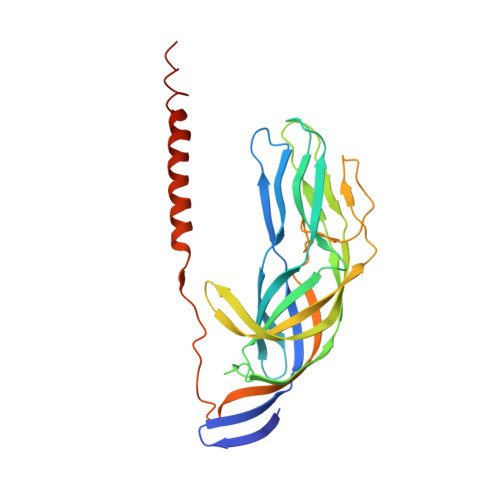
1SLQ

Crystal structure of the trimeric state of the rhesus rotavirus VP4 membrane interaction domain, VP5CT


Experimental Data Snapshot

  • Method: X-RAY DIFFRACTION
  • Resolution: 3.20 Å
  • R-Value Free: 0.338 
  • R-Value Work: 0.308 
  • R-Value Observed: 0.310 

wwPDB Validation   3D Report Full Report


This is version 1.3 of the entry. See complete history


Literature

Structural rearrangements in the membrane penetration protein of a non-enveloped virus.

Dormitzer, P.R.Nason, E.B.Prasad, B.V.Harrison, S.C.

(2004) Nature 430: 1053-1058

  • DOI: https://doi.org/10.1038/nature02836
  • Primary Citation of Related Structures:  
    1SLQ

  • PubMed Abstract: 

    Non-enveloped virus particles (those that lack a lipid-bilayer membrane) must breach the membrane of a target host cell to gain access to its cytoplasm. So far, the molecular mechanism of this membrane penetration step has resisted structural analysis. The spike protein VP4 is a principal component in the entry apparatus of rotavirus, a non-enveloped virus that causes gastroenteritis and kills 440,000 children each year. Trypsin cleavage of VP4 primes the virus for entry by triggering a rearrangement that rigidifies the VP4 spikes. We have determined the crystal structure, at 3.2 A resolution, of the main part of VP4 that projects from the virion. The crystal structure reveals a coiled-coil stabilized trimer. Comparison of this structure with the two-fold clustered VP4 spikes in a approximately 12 A resolution image reconstruction from electron cryomicroscopy of trypsin-primed virions shows that VP4 also undergoes a second rearrangement, in which the oligomer reorganizes and each subunit folds back on itself, translocating a potential membrane-interaction peptide from one end of the spike to the other. This rearrangement resembles the conformational transitions of membrane fusion proteins of enveloped viruses.


  • Organizational Affiliation

    Department of Pediatrics, Harvard Medical School, and the Laboratory of Molecular Medicine, Children's Hospital, Boston, Massachusetts 02115, USA. dormitze@crystal.harvard.edu


Macromolecules
Find similar proteins by:  (by identity cutoff)  |  3D Structure
Entity ID: 1
MoleculeChains Sequence LengthOrganismDetailsImage
VP4
A, B, C, D, E
A, B, C, D, E, F
278Simian rotavirus A strain RRVMutation(s): 0 
Gene Names: gene segment 4
UniProt
Find proteins for P12473 (Rotavirus A (strain RVA/Monkey/United States/RRV/1975/G3P5B[3]))
Explore P12473 
Go to UniProtKB:  P12473
Entity Groups  
Sequence Clusters30% Identity50% Identity70% Identity90% Identity95% Identity100% Identity
UniProt GroupP12473
Sequence Annotations
Expand
  • Reference Sequence
Experimental Data & Validation

Experimental Data

  • Method: X-RAY DIFFRACTION
  • Resolution: 3.20 Å
  • R-Value Free: 0.338 
  • R-Value Work: 0.308 
  • R-Value Observed: 0.310 
  • Space Group: P 42 2 2
Unit Cell:
Length ( Å )Angle ( ˚ )
a = 244.843α = 90
b = 244.843β = 90
c = 104.778γ = 90
Software Package:
Software NamePurpose
HKL-2000data collection
SCALEPACKdata scaling
MLPHAREphasing
CNSrefinement
HKL-2000data reduction

Structure Validation

View Full Validation Report


Entry History 

Deposition Data

Revision History  (Full details and data files)

  • Version 1.0: 2004-08-31
    Type: Initial release
  • Version 1.1: 2008-04-29
    Changes: Version format compliance
  • Version 1.2: 2011-07-13
    Changes: Version format compliance
  • Version 1.3: 2024-02-14
    Changes: Data collection, Database references