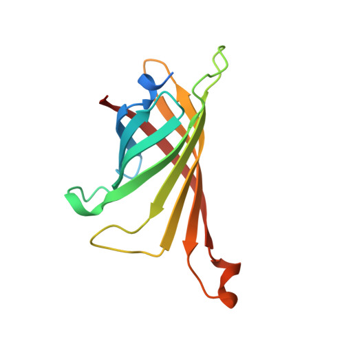
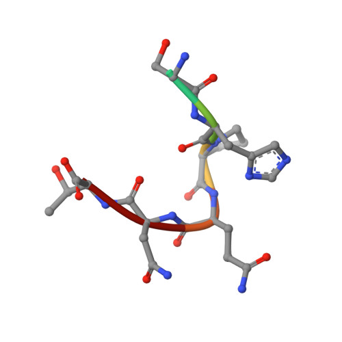
Binding to protein targets of peptidic leads discovered by phage display: crystal structures of streptavidin-bound linear and cyclic peptide ligands containing the HPQ sequence
Katz, B.A.(1995) Biochemistry 34: 15421-15429
- PubMed: 7492542
- DOI: https://doi.org/10.1021/bi00047a005
- Primary Citation of Related Structures:
1SLD, 1SLE, 1SLF, 1SLG - PubMed Abstract:
The streptavidin-bound crystal structures of two disulfide-bridge cyclic peptides (cyclo-Ac-[CHPQGPPC]-NH2 and cyclo-Ac-[CHPQFC]-NH2) and of a linear peptide (FSHPQNT) were determined, as well as the structure of apostreptavidin (streptavidin-sulfate). Both the linear and disulfide-bridged cyclic peptides studied share a common HPQ conformation and make common interactions with streptavidin, although significant differences in structures and interactions occur for flanking residues among the complexes. The conformation of the linear peptide in the crystal structure of streptavidin-FSHPQNT was found to differ from that in the same complex published [Weber, P. C., Pantoliano, M. W., & Thompson, L. D. (1992) Biochemistry 31, 9350-9354]. In the present investigation, the HPQNT portion of the ligand is well-defined with some density defining the Phe, whereas in the investigation of Weber et al. only the HPQ segment of the bound peptide could be interpreted. Both bound cyclic peptides adopt a beta-turn involving an H-bond between the His main chain carbonyl and the main chain amide NH of the i+3 residue. In the streptavidin-bound cyclo-Ac-[CHPQFC]-NH2 structure, there is an additional H-bond, indicative of alpha-helix, between the main chain His carbonyl and the main chain C-terminal Cys amide NH group. Binding interactions for both cyclic and linear peptides include direct H-bonds, H-bonds mediated by tightly bound water molecules, and hydrophobic interactions. The above structures and that of streptavidin-biotin in the literature are compared and discussed in the context of structure-based ligand design.
- Arris Pharmaceutical Corporation, South San Francisco, California 94080, USA.
Organizational Affiliation:


