1SHW | pdb_00001shw

EphB2 / EphrinA5 Complex Structure


Experimental Data Snapshot

  • Method: X-RAY DIFFRACTION
  • Resolution: 2.20 Å
  • R-Value Free: 
    0.305 (Depositor) 
  • R-Value Work: 
    0.258 (Depositor) 

Starting Model: experimental
View more details

wwPDB Validation 3D Report Full Report

Validation slider image for 1SHW

This is version 2.2 of the entry. See complete history. 

Literature

Repelling class discrimination: ephrin-A5 binds to and activates EphB2 receptor signaling

Himanen, J.P., Chumley, M.J., Lackmann, M., Li, C., Barton, W.A., Jeffrey, P.D., Vearing, C., Geleick, D., Feldheim, D.A., Boyd, A.W., Henkemeyer, M., Nikolov, D.B.

(2004) Nat Neurosci 7: 501-509

  • DOI: https://doi.org/10.1038/nn1237
  • Primary Citation Related Structures: 
    1SHW

  • PubMed Abstract: 

    The interactions between Eph receptor tyrosine kinases and their ephrin ligands regulate cell migration and axon pathfinding. The EphA receptors are generally thought to become activated by ephrin-A ligands, whereas the EphB receptors interact with ephrin-B ligands. Here we show that two of the most widely studied of these molecules, EphB2 and ephrin-A5, which have never been described to interact with each other, do in fact bind one another with high affinity. Exposure of EphB2-expressing cells to ephrin-A5 leads to receptor clustering, autophosphorylation and initiation of downstream signaling. Ephrin-A5 induces EphB2-mediated growth cone collapse and neurite retraction in a model system. We further show, using X-ray crystallography, that the ephrin-A5-EphB2 complex is a heterodimer and is architecturally distinct from the tetrameric EphB2-ephrin-B2 structure. The structural data reveal the molecular basis for EphB2-ephrin-A5 signaling and provide a framework for understanding the complexities of functional interactions and crosstalk between A- and B-subclass Eph receptors and ephrins.


  • Organizational Affiliation: 
    • Structural Biology Program, Memorial Sloan-Kettering Cancer Center, New York, New York 10021, USA.

Macromolecule Content 

  • Total Structure Weight: 37.9 kDa 
  • Atom Count: 2,880 
  • Modeled Residue Count: 319 
  • Deposited Residue Count: 319 
  • Unique protein chains: 2

Macromolecules

Find similar proteins by:|  3D Structure
Entity ID: 1
MoleculeChains  Sequence LengthOrganismDetailsImage
Ephrin-A5138Mus musculusMutation(s): 0 
Gene Names: EFNA5, EPLG7, LERK7, EPL7
UniProt & NIH Common Fund Data Resources
Find proteins for O08543 (Mus musculus)
Explore O08543 
Go to UniProtKB:  O08543
IMPC:  MGI:107444
Entity Groups
Sequence Clusters30% Identity50% Identity70% Identity90% Identity95% Identity100% Identity
UniProt GroupO08543
Glycosylation
Glycosylation Sites: 1Go to GlyGen: O08543-1
Sequence Annotations
Expand
Reference Sequence
Find similar proteins by:|  3D Structure
Entity ID: 2
MoleculeChains  Sequence LengthOrganismDetailsImage
Ephrin type-B receptor 2181Mus musculusMutation(s): 0 
Gene Names: EPHB2, EPTH3, NUK, SEK3
EC: 2.7.10.1
UniProt & NIH Common Fund Data Resources
Find proteins for P54763 (Mus musculus)
Explore P54763 
Go to UniProtKB:  P54763
IMPC:  MGI:99611
Entity Groups
Sequence Clusters30% Identity50% Identity70% Identity90% Identity95% Identity100% Identity
UniProt GroupP54763
Sequence Annotations
Expand
Reference Sequence

Oligosaccharides

Help  
Entity ID: 3
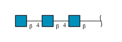
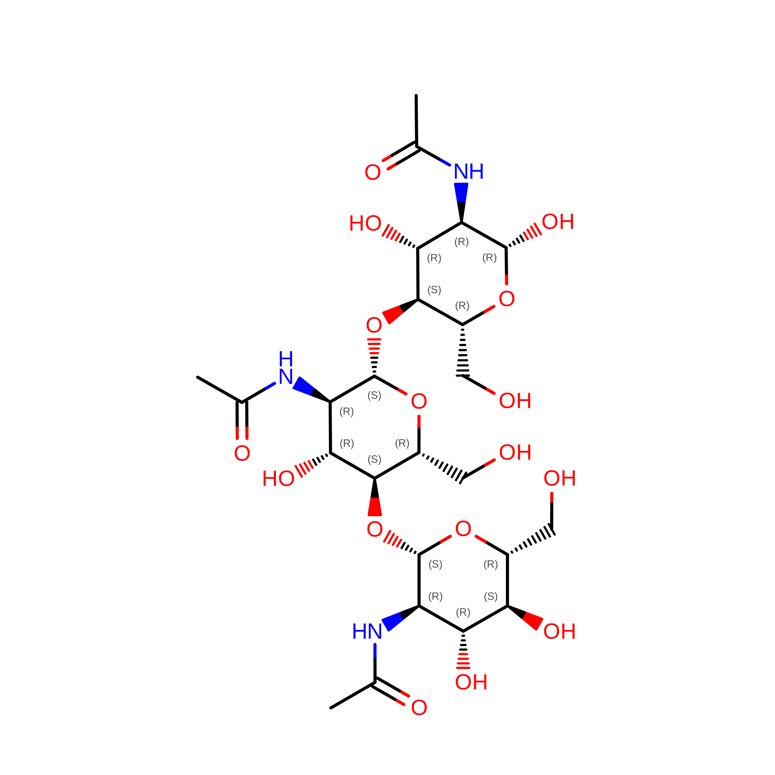
MoleculeChains Length2D Diagram GlycosylationD Interactions
2-acetamido-2-deoxy-beta-D-glucopyranose-(1-4)-2-acetamido-2-deoxy-beta-D-glucopyranose-(1-4)-2-acetamido-2-deoxy-beta-D-glucopyranose
C
3N-Glycosylation
Glycosylation Resources
GlyTouCan: G47362BJ
GlyCosmos: G47362BJ
GlyGen: G47362BJ

Small Molecules

Ligands 1 Unique
IDChains Name / Formula / InChI Key2D Diagram3D Interactions
ZN

Query on ZN


Download:Ideal Coordinates CCD File
D [auth A]ZINC ION
Zn
PTFCDOFLOPIGGS-UHFFFAOYSA-N

Biologically Interesting Molecules (External Reference) 

1 Unique

Experimental Data & Validation

Experimental Data

  • Method: X-RAY DIFFRACTION
  • Resolution: 2.20 Å
  • R-Value Free:  0.305 (Depositor) 
  • R-Value Work:  0.258 (Depositor) 
Space Group: P 21 21 21
Unit Cell:
Length ( Å )Angle ( ˚ )
a = 37.89α = 90
b = 88.97β = 90
c = 122.81γ = 90
Software Package:
Software NamePurpose
DENZOdata reduction
SCALEPACKdata scaling
AMoREphasing
CNSrefinement

Structure Validation

View Full Validation Report


Entry History 

Deposition Data

Revision History  (Full details and data files)

  • Version 1.0: 2004-05-18
    Type: Initial release
  • Version 1.1: 2008-04-29
    Changes: Version format compliance
  • Version 1.2: 2011-07-13
    Changes: Non-polymer description, Version format compliance
  • Version 2.0: 2020-07-29
    Type: Remediation
    Reason: Carbohydrate remediation
    Changes: Advisory, Atomic model, Data collection, Derived calculations, Structure summary
  • Version 2.1: 2024-04-03
    Changes: Advisory, Data collection, Database references, Refinement description, Structure summary
  • Version 2.2: 2024-10-30
    Changes: Structure summary