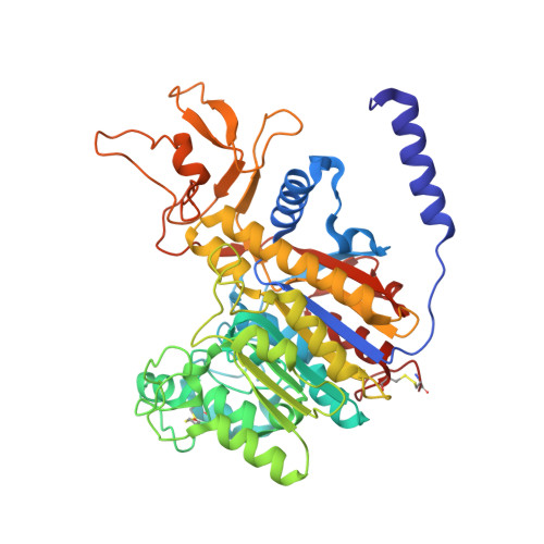
1SHN

Crystal structure of shrimp alkaline phosphatase with phosphate bound


Experimental Data Snapshot

  • Method: X-RAY DIFFRACTION
  • Resolution: 2.15 Å
  • R-Value Free: 0.218 
  • R-Value Work: 0.186 
  • R-Value Observed: 0.188 

wwPDB Validation   3D Report Full Report


Ligand Structure Quality Assessment 


This is version 1.3 of the entry. See complete history


Literature

Ligand-binding and metal-exchange crystallographic studies on shrimp alkaline phosphatase.

de Backer, M.M.McSweeney, S.Lindley, P.F.Hough, E.

(2004) Acta Crystallogr D Biol Crystallogr 60: 1555-1561

  • DOI: https://doi.org/10.1107/S0907444904015628
  • Primary Citation of Related Structures:  
    1SHN, 1SHQ

  • PubMed Abstract: 

    Alkaline phosphatases (APs) are homodimeric metalloenzymes that catalyze the hydrolysis and transphosphorylation of phosphate monoesters. Each monomer contains a metal-binding triad that for optimal activity is usually occupied by two zinc ions and one magnesium ion. The recently determined crystal structure of cold-active shrimp alkaline phosphatase (SAP) was, however, fully occupied by zinc ions. This paper describes a metal-exchange experiment in which the zinc ion in one binding site (referred to as the M3 site) is replaced by magnesium. Crystal structures revealed a concomitant structural change: the metal exchange causes movement of a ligating histidine into a conformation in which it does not coordinate to the metal ion. The M3 site is relevant to catalysis: its occupation by magnesium is postulated to favour catalysis and it has been suggested to be a regulatory site for other APs. Further crystallographic studies show that ligand binding can induce a conformational change of an active-site arginine from a 'non-docked' (non-interacting) to a 'docked' conformation (interacting with the ligand). The first conformation has only been observed in SAP, while the latter is common in available AP structures. The observation that the arginine does not always bind the substrate may explain the increased catalytic efficiency that is generally observed for cold-active enzymes.


  • Organizational Affiliation

    European Synchrotron Radiation Facility, France.


Macromolecules
Find similar proteins by:  (by identity cutoff)  |  3D Structure
Entity ID: 1
MoleculeChains Sequence LengthOrganismDetailsImage
alkaline phosphatase
A, B
478Pandalus borealisMutation(s): 0 
EC: 3.1.3.1
UniProt
Find proteins for Q9BHT8 (Pandalus borealis)
Explore Q9BHT8 
Go to UniProtKB:  Q9BHT8
Entity Groups  
Sequence Clusters30% Identity50% Identity70% Identity90% Identity95% Identity100% Identity
UniProt GroupQ9BHT8
Glycosylation
Glycosylation Sites: 1
Sequence Annotations
Expand
  • Reference Sequence
Small Molecules
Ligands 4 Unique
IDChains Name / Formula / InChI Key2D Diagram3D Interactions
NAG
Query on NAG

Download Ideal Coordinates CCD File 
C [auth A],
I [auth B]
2-acetamido-2-deoxy-beta-D-glucopyranose
C8 H15 N O6
OVRNDRQMDRJTHS-FMDGEEDCSA-N
SO4
Query on SO4

Download Ideal Coordinates CCD File 
H [auth A],
N [auth B]
SULFATE ION
O4 S
QAOWNCQODCNURD-UHFFFAOYSA-L
PO4
Query on PO4

Download Ideal Coordinates CCD File 
G [auth A],
M [auth B]
PHOSPHATE ION
O4 P
NBIIXXVUZAFLBC-UHFFFAOYSA-K
ZN
Query on ZN

Download Ideal Coordinates CCD File 
D [auth A]
E [auth A]
F [auth A]
J [auth B]
K [auth B]
D [auth A],
E [auth A],
F [auth A],
J [auth B],
K [auth B],
L [auth B]
ZINC ION
Zn
PTFCDOFLOPIGGS-UHFFFAOYSA-N
Experimental Data & Validation

Experimental Data

  • Method: X-RAY DIFFRACTION
  • Resolution: 2.15 Å
  • R-Value Free: 0.218 
  • R-Value Work: 0.186 
  • R-Value Observed: 0.188 
  • Space Group: P 43 21 2
Unit Cell:
Length ( Å )Angle ( ˚ )
a = 170.676α = 90
b = 170.677β = 90
c = 83.925γ = 90
Software Package:
Software NamePurpose
REFMACrefinement
DENZOdata reduction
SCALEPACKdata scaling
MLPHAREphasing

Structure Validation

View Full Validation Report


Ligand Structure Quality Assessment 


Entry History 

Deposition Data

Revision History  (Full details and data files)

  • Version 1.0: 2004-08-31
    Type: Initial release
  • Version 1.1: 2008-04-29
    Changes: Version format compliance
  • Version 1.2: 2011-07-13
    Changes: Non-polymer description, Version format compliance
  • Version 1.3: 2020-07-29
    Type: Remediation
    Reason: Carbohydrate remediation
    Changes: Data collection, Database references, Derived calculations, Structure summary