1SGQ | pdb_00001sgq

GLY 18 VARIANT OF TURKEY OVOMUCOID INHIBITOR THIRD DOMAIN COMPLEXED WITH STREPTOMYCES GRISEUS PROTEINASE B


Experimental Data Snapshot

  • Method: X-RAY DIFFRACTION
  • Resolution: 1.90 Å
  • R-Value Observed: 
    0.131 (Depositor) 

wwPDB Validation 3D Report Full Report

Validation slider image for 1SGQ

This is version 1.4 of the entry. See complete history

Literature

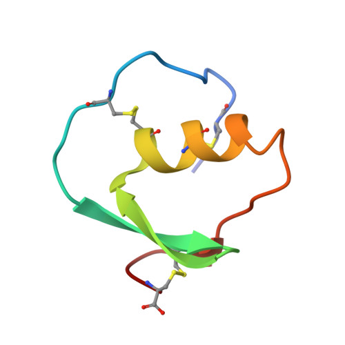
Water molecules participate in proteinase-inhibitor interactions: crystal structures of Leu18, Ala18, and Gly18 variants of turkey ovomucoid inhibitor third domain complexed with Streptomyces griseus proteinase B.

Huang, K.Lu, W.Anderson, S.Laskowski Jr., M.James, M.N.

(1995) Protein Sci 4: 1985-1997

  • DOI: https://doi.org/10.1002/pro.5560041004
  • Primary Citation Related Structures: 
    1SGP, 1SGQ, 1SGR, 2SGE

  • PubMed Abstract: 

    Crystal structures of the complexes of Streptomyces griseus proteinase B (SGPB) with three P1 variants of turkey ovomucoid inhibitor third domain (OMTKY3), Leu18, Ala18, and Gly18, have been determined and refined to high resolution. Comparisons among these structures and of each with native, uncomplexed SGPB reveal that each complex features a unique solvent structure in the S1 binding pocket. The number and relative positions of water molecules bound in the S1 binding pocket vary according to the size of the side chain of the P1 residue. Water molecules in the S1 binding pocket of SGPB are redistributed in response to the complex formation, probably to optimize hydrogen bonds between the enzyme and the inhibitor. There are extensive water-mediated hydrogen bonds in the interfaces of the complexes. In all complexes, Asn 36 of OMTKY3 participates in forming hydrogen bonds, via water molecules, with residues lining the S1 binding pocket of SGPB. For a homologous series of aliphatic straight side chains, Gly18, Ala18, Abu18, Ape18, and Ahp18 variants, the binding free energy is a linear function of the hydrophobic surface area buried in the interface of the corresponding complexes. The resulting constant of proportionality is 34.1 cal mol-1 A-2. These structures confirm that the binding of OMTKY3 to the preformed S1 pocket in SGPB involves no substantial structural disturbances that commonly occur in the site-directed mutagenesis studies of interior residues in other proteins, thus providing one of the most reliable assessments of the contribution of the hydrophobic effect to protein-complex stability.


  • Organizational Affiliation
    • Department of Biochemistry, University of Alberta, Edmonton, Canada.

Macromolecule Content 

  • Total Structure Weight: 24.29 kDa 
  • Atom Count: 1,866 
  • Modeled Residue Count: 236 
  • Deposited Residue Count: 236 
  • Unique protein chains: 2

Macromolecules

Find similar proteins by:|  3D Structure
Entity ID: 1
MoleculeChains  Sequence LengthOrganismDetailsImage
STREPTOMYCES GRISEUS PROTEINASE BA [auth E]185Streptomyces griseusMutation(s): 0 
EC: 3.4.21.81
UniProt
Find proteins for P00777 (Streptomyces griseus)
Explore P00777 
Go to UniProtKB:  P00777
Entity Groups
Sequence Clusters30% Identity50% Identity70% Identity90% Identity95% Identity100% Identity
UniProt GroupP00777
Sequence Annotations
Expand
Reference Sequence
Find similar proteins by:|  3D Structure
Entity ID: 2
MoleculeChains  Sequence LengthOrganismDetailsImage
TURKEY OVOMUCOID INHIBITORB [auth I]51Meleagris gallopavoMutation(s): 0 
UniProt
Find proteins for P68390 (Meleagris gallopavo)
Explore P68390 
Go to UniProtKB:  P68390
Entity Groups
Sequence Clusters30% Identity50% Identity70% Identity90% Identity95% Identity100% Identity
UniProt GroupP68390
Sequence Annotations
Expand
Reference Sequence

Small Molecules

Ligands 1 Unique
IDChains Name / Formula / InChI Key2D Diagram3D Interactions
PO4

Query on PO4


Download:Ideal Coordinates CCD File
C [auth E]PHOSPHATE ION
O4 P
NBIIXXVUZAFLBC-UHFFFAOYSA-K

Experimental Data & Validation

Experimental Data

  • Method: X-RAY DIFFRACTION
  • Resolution: 1.90 Å
  • R-Value Observed: 0.131 (Depositor) 
Space Group: P 1 21 1
Unit Cell:
Length ( Å )Angle ( ˚ )
a = 45.48α = 90
b = 54.68β = 118.99
c = 45.55γ = 90
Software Package:
Software NamePurpose
BIOMOLdata collection
TNTrefinement
BIOMOLdata reduction

Structure Validation

View Full Validation Report


Entry History 

Deposition Data

Revision History  (Full details and data files)

  • Version 1.0: 1995-10-15
    Type: Initial release
  • Version 1.1: 2008-03-03
    Changes: Version format compliance
  • Version 1.2: 2011-07-13
    Changes: Version format compliance
  • Version 1.3: 2017-11-29
    Changes: Derived calculations, Other
  • Version 1.4: 2024-11-06
    Changes: Data collection, Database references, Derived calculations, Structure summary