1SFK

Core (C) protein from West Nile Virus, subtype Kunjin


Experimental Data Snapshot

  • Method: X-RAY DIFFRACTION
  • Resolution: 3.20 Å
  • R-Value Free: 0.311 
  • R-Value Work: 0.249 
  • R-Value Observed: 0.252 

wwPDB Validation   3D Report Full Report


This is version 1.2 of the entry. See complete history


Literature

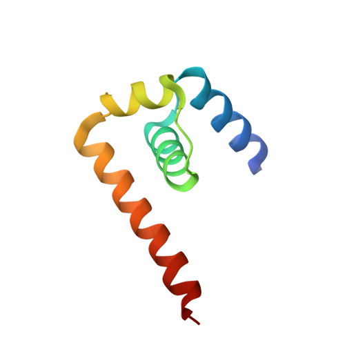
West nile virus core protein; tetramer structure and ribbon formation

Dokland, T.Walsh, M.Mackenzie, J.M.Khromykh, A.A.Ee, K.-H.Wang, S.

(2004) Structure 12: 1157-1163

  • DOI: 10.1016/j.str.2004.04.024
  • Primary Citation of Related Structures:  
    1SFK

  • PubMed Abstract: 
  • We have determined the crystal structure of the core (C) protein from the Kunjin subtype of West Nile virus (WNV), closely related to the NY99 strain of WNV, currently a major health threat in the U.S. WNV is a member of the Flaviviridae family of enveloped RNA viruses that contains many important human pathogens ...

    We have determined the crystal structure of the core (C) protein from the Kunjin subtype of West Nile virus (WNV), closely related to the NY99 strain of WNV, currently a major health threat in the U.S. WNV is a member of the Flaviviridae family of enveloped RNA viruses that contains many important human pathogens. The C protein is associated with the RNA genome and forms the internal core which is surrounded by the envelope in the virion. The C protein structure contains four alpha helices and forms dimers that are organized into tetramers. The tetramers form extended filamentous ribbons resembling the stacked alpha helices seen in HEAT protein structures.


    Organizational Affiliation

    Institute of Molecular and Cell Biology, Singapore, Republic of Singapore. dokland@uab.edu


Macromolecules
Find similar proteins by:  (by identity cutoff)  |  3D Structure
Entity ID: 1
MoleculeChainsSequence LengthOrganismDetailsImage
Core protein
A, B, C, D, E, F, G, H
76Kunjin virus (STRAIN MRM61C)Mutation(s): 0 
EC: 3.4.21.91 (UniProt), 3.6.1.15 (UniProt), 3.6.4.13 (UniProt), 2.1.1.56 (UniProt), 2.1.1.57 (UniProt), 2.7.7.48 (UniProt)
UniProt
Find proteins for P14335 (Kunjin virus (strain MRM61C))
Explore P14335 
Go to UniProtKB:  P14335
Entity Groups  
Sequence Clusters30% Identity50% Identity70% Identity90% Identity95% Identity100% Identity
UniProt GroupP14335
Protein Feature View
Expand
  • Reference Sequence
Small Molecules
Ligands 4 Unique
IDChainsName / Formula / InChI Key2D Diagram3D Interactions
PG4
Query on PG4

Download Ideal Coordinates CCD File 
L [auth A],
O [auth D],
R [auth F],
U [auth G]
TETRAETHYLENE GLYCOL
C8 H18 O5
UWHCKJMYHZGTIT-UHFFFAOYSA-N
 Ligand Interaction
PO4
Query on PO4

Download Ideal Coordinates CCD File 
K [auth A]PHOSPHATE ION
O4 P
NBIIXXVUZAFLBC-UHFFFAOYSA-K
 Ligand Interaction
CA
Query on CA

Download Ideal Coordinates CCD File 
I [auth A],
M [auth D],
P [auth F],
S [auth G]
CALCIUM ION
Ca
BHPQYMZQTOCNFJ-UHFFFAOYSA-N
 Ligand Interaction
CL
Query on CL

Download Ideal Coordinates CCD File 
J [auth A],
N [auth D],
Q [auth F],
T [auth G]
CHLORIDE ION
Cl
VEXZGXHMUGYJMC-UHFFFAOYSA-M
 Ligand Interaction
Experimental Data & Validation

Experimental Data

  • Method: X-RAY DIFFRACTION
  • Resolution: 3.20 Å
  • R-Value Free: 0.311 
  • R-Value Work: 0.249 
  • R-Value Observed: 0.252 
  • Space Group: I 41
Unit Cell:
Length ( Å )Angle ( ˚ )
a = 85.655α = 90
b = 85.655β = 90
c = 214.384γ = 90
Software Package:
Software NamePurpose
REFMACrefinement
HKL-2000data reduction
SCALEPACKdata scaling
SOLVEphasing

Structure Validation

View Full Validation Report


Entry History 

Deposition Data

Revision History  (Full details and data files)

  • Version 1.0: 2004-08-09
    Type: Initial release
  • Version 1.1: 2008-04-29
    Changes: Version format compliance
  • Version 1.2: 2011-07-13
    Changes: Advisory, Derived calculations, Version format compliance