1SCH

PEANUT PEROXIDASE


Experimental Data Snapshot

  • Method: X-RAY DIFFRACTION
  • Resolution: 2.56 Å
  • R-Value Free: 0.262 
  • R-Value Work: 0.199 
  • R-Value Observed: 0.199 

wwPDB Validation   3D Report Full Report


This is version 2.1 of the entry. See complete history


Literature

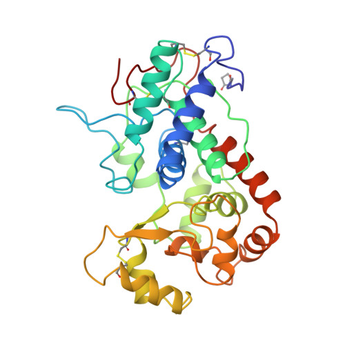
The crystal structure of peanut peroxidase.

Schuller, D.J.Ban, N.Huystee, R.B.McPherson, A.Poulos, T.L.

(1996) Structure 4: 311-321

  • DOI: https://doi.org/10.1016/s0969-2126(96)00035-4
  • Primary Citation of Related Structures:  
    1SCH

  • PubMed Abstract: 
  • Peroxidases catalyze a wide variety of peroxide-dependent oxidations. Based on sequence alignments, heme peroxidases have been divided into three classes. Crystal structures are available for peroxidases of classes I and II, but until now no structure has been determined for class III, the classical extracellular plant peroxidases ...

    Peroxidases catalyze a wide variety of peroxide-dependent oxidations. Based on sequence alignments, heme peroxidases have been divided into three classes. Crystal structures are available for peroxidases of classes I and II, but until now no structure has been determined for class III, the classical extracellular plant peroxidases.


    Organizational Affiliation

    Department of Molecular Biology & Biochemistry, University of California, Irvine, CA 92717-3900, USA.


Macromolecules
Find similar proteins by:  (by identity cutoff)  |  3D Structure
Entity ID: 1
MoleculeChainsSequence LengthOrganismDetailsImage
PEANUT PEROXIDASE, MAJOR CATIONIC ISOZYME
A, B
294Arachis hypogaeaMutation(s): 0 
Gene Names: PNC1
EC: 1.11.1.7
UniProt
Find proteins for P22195 (Arachis hypogaea)
Explore P22195 
Go to UniProtKB:  P22195
Entity Groups  
Sequence Clusters30% Identity50% Identity70% Identity90% Identity95% Identity100% Identity
UniProt GroupP22195
Protein Feature View
Expand
  • Reference Sequence
Small Molecules
Ligands 3 Unique
IDChainsName / Formula / InChI Key2D Diagram3D Interactions
HEM
Query on HEM

Download Ideal Coordinates CCD File 
E [auth A],
I [auth B]
PROTOPORPHYRIN IX CONTAINING FE
C34 H32 Fe N4 O4
KABFMIBPWCXCRK-RGGAHWMASA-L
 Ligand Interaction
NAG
Query on NAG

Download Ideal Coordinates CCD File 
F [auth B]2-acetamido-2-deoxy-beta-D-glucopyranose
C8 H15 N O6
OVRNDRQMDRJTHS-FMDGEEDCSA-N
 Ligand Interaction
CA
Query on CA

Download Ideal Coordinates CCD File 
C [auth A],
D [auth A],
G [auth B],
H [auth B]
CALCIUM ION
Ca
BHPQYMZQTOCNFJ-UHFFFAOYSA-N
 Ligand Interaction
Modified Residues  1 Unique
IDChainsTypeFormula2D DiagramParent
PCA
Query on PCA
A, B
L-PEPTIDE LINKINGC5 H7 N O3GLN
Experimental Data & Validation

Experimental Data

  • Method: X-RAY DIFFRACTION
  • Resolution: 2.56 Å
  • R-Value Free: 0.262 
  • R-Value Work: 0.199 
  • R-Value Observed: 0.199 
  • Space Group: P 21 21 21
Unit Cell:
Length ( Å )Angle ( ˚ )
a = 48.1α = 90
b = 97.2β = 90
c = 146.2γ = 90
Software Package:
Software NamePurpose
HOWARDdata collection
NIELSENdata collection
X-PLORmodel building
X-PLORrefinement
XENGENdata reduction
NIELSENdata reduction
XUONG)data reduction
X-PLORphasing

Structure Validation

View Full Validation Report


Entry History 

Deposition Data

Revision History  (Full details and data files)

  • Version 1.0: 1996-07-11
    Type: Initial release
  • Version 1.1: 2008-03-24
    Changes: Version format compliance
  • Version 1.2: 2011-07-13
    Changes: Non-polymer description, Version format compliance
  • Version 2.0: 2019-12-25
    Changes: Derived calculations, Other, Polymer sequence
  • Version 2.1: 2020-07-29
    Type: Remediation
    Reason: Carbohydrate remediation
    Changes: Data collection, Derived calculations, Structure summary