1S8N | pdb_00001s8n

Crystal structure of Rv1626 from Mycobacterium tuberculosis


Experimental Data Snapshot

  • Method: X-RAY DIFFRACTION
  • Resolution: 1.48 Å
  • R-Value Free: 
    0.228 (Depositor), 0.262 (DCC) 
  • R-Value Work: 
    0.203 (Depositor), 0.244 (DCC) 
  • R-Value Observed: 
    0.204 (Depositor) 

Starting Model: experimental
View more details

wwPDB Validation 3D Report Full Report

Validation slider image for 1S8N

This is version 1.3 of the entry. See complete history

Literature

The Crystal and Solution Structure of a Putative Transcriptional Antiterminator from Mycobacterium tuberculosis.

Morth, J.P.Feng, V.Perry, L.J.Svergun, D.I.Tucker, P.A.

(2004) Structure 12: 1595-1605

  • DOI: https://doi.org/10.1016/j.str.2004.06.018
  • Primary Citation Related Structures: 
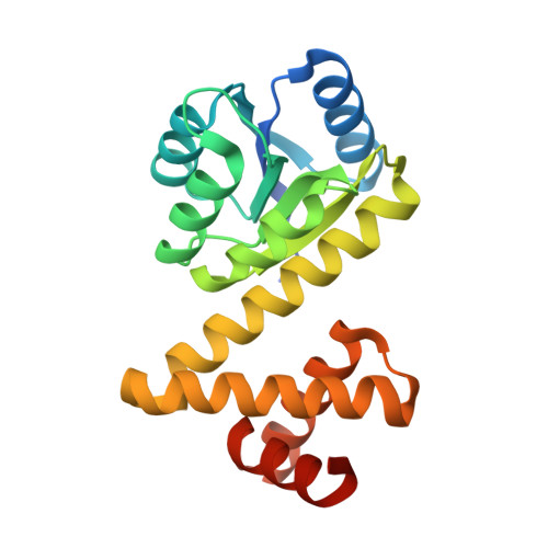
    1S8N, 1SD5

  • PubMed Abstract: 

    We describe the crystal structure of Rv1626 from Mycobacterium tuberculosis at 1.48 A resolution and the corresponding solution structure determined from small angle X-ray scattering. The N-terminal domain shows structural homology to the receiver domains found in bacterial two-component systems. The C-terminal domain has high structural homology to a recently discovered RNA binding domain involved in transcriptional antitermination. The molecule in solution was found to be monomeric as it is in the crystal, but in solution it undergoes a conformational change that is triggered by changes in ionic strength. This is the first structure that links the phosphorylation cascade of the two-component systems with the antitermination event in the transcriptional machinery. Rv1626 belongs to a family of proteins, which we propose calling phosphorylation-dependent transcriptional antitermination regulators, so far only found in bacteria, and includes NasT, a protein from the assimilatory nitrate/nitrite reductase operon of Azetobacter vinelandii.


  • Organizational Affiliation
    • EMBL Hamburg Outstation, c/o DESY, Notkestrasse 85, Germany.

Macromolecule Content 

  • Total Structure Weight: 22.74 kDa 
  • Atom Count: 1,736 
  • Modeled Residue Count: 190 
  • Deposited Residue Count: 205 
  • Unique protein chains: 1

Macromolecules

Find similar proteins by:|  3D Structure
Entity ID: 1
MoleculeChains  Sequence LengthOrganismDetailsImage
putative antiterminator205Mycobacterium tuberculosis H37RvMutation(s): 0 
Gene Names: rv1626
UniProt
Find proteins for P9WGM3 (Mycobacterium tuberculosis (strain ATCC 25618 / H37Rv))
Explore P9WGM3 
Go to UniProtKB:  P9WGM3
Entity Groups
Sequence Clusters30% Identity50% Identity70% Identity90% Identity95% Identity100% Identity
UniProt GroupP9WGM3
Sequence Annotations
Expand
Reference Sequence

Small Molecules

Ligands 1 Unique
IDChains Name / Formula / InChI Key2D Diagram3D Interactions
AZI

Query on AZI


Download:Ideal Coordinates CCD File
B [auth A]AZIDE ION
N3
IVRMZWNICZWHMI-UHFFFAOYSA-N

Experimental Data & Validation

Experimental Data

  • Method: X-RAY DIFFRACTION
  • Resolution: 1.48 Å
  • R-Value Free:  0.228 (Depositor), 0.262 (DCC) 
  • R-Value Work:  0.203 (Depositor), 0.244 (DCC) 
  • R-Value Observed: 0.204 (Depositor) 
Space Group: P 43 21 2
Unit Cell:
Length ( Å )Angle ( ˚ )
a = 44.836α = 90
b = 44.836β = 90
c = 180.626γ = 90
Software Package:
Software NamePurpose
REFMACrefinement
DENZOdata reduction
CCP4data scaling
MOLREPphasing

Structure Validation

View Full Validation Report


Entry History 

Deposition Data

Revision History  (Full details and data files)

  • Version 1.0: 2004-09-21
    Type: Initial release
  • Version 1.1: 2008-04-29
    Changes: Version format compliance
  • Version 1.2: 2011-07-13
    Changes: Source and taxonomy, Version format compliance
  • Version 1.3: 2023-08-23
    Changes: Data collection, Database references, Derived calculations, Refinement description