1S66 | pdb_00001s66

Crystal structure of heme domain of direct oxygen sensor from E. coli


Experimental Data Snapshot

  • Method: X-RAY DIFFRACTION
  • Resolution: 1.80 Å
  • R-Value Free: 
    0.224 (Depositor) 
  • R-Value Work: 
    0.189 (Depositor) 
  • R-Value Observed: 
    0.234 (Depositor) 

wwPDB Validation 3D Report Full Report

Validation slider image for 1S66

This is version 1.3 of the entry. See complete history

Literature

Insights into signal transduction involving PAS domain oxygen-sensing heme proteins from the X-ray crystal structure of Escherichia coli Dos heme domain (Ec DosH)

Park, H.J.Suquet, C.Satterlee, J.D.Kang, C.H.

(2004) Biochemistry 43: 2738-2746

  • DOI: https://doi.org/10.1021/bi035980p
  • Primary Citation Related Structures: 
    1S66, 1S67

  • PubMed Abstract: 

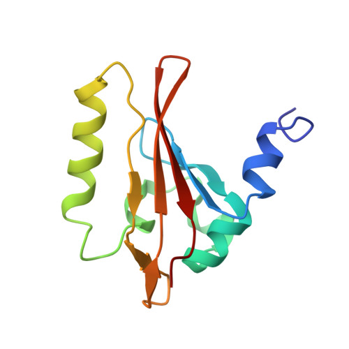
    The X-ray crystal structure of the Escherichia coli (Ec) direct oxygen sensor heme domain (Ec DosH) has been solved to 1.8 A using Fe multiple-wavelength anomalous dispersion (MAD), and the positions of Met95 have been confirmed by selenomethionine ((Se)Met) MAD. Ec DosH is the sensing part of a larger two-domain sensing/signaling protein, in which the signaling domain has phosphodiesterase activity. The asymmetric unit of the crystal lattice contains a dimer comprised of two differently ligated heme domain monomers. Except for the heme ligands, the monomer heme domains are identical. In one monomer, the heme is ligated by molecular oxygen (O(2)), while in the other monomer, an endogenous Met95 with S --> Fe ligation replaces the exogenous O(2) ligand. In both heme domains, the proximal ligand is His77. Analysis of these structures reveals sizable ligand-dependent conformational changes in the protein chain localized in the FG turn, the G(beta)-strand, and the HI turn. These changes provide insight to the mechanism of signal propagation within the heme domain following initiation due to O(2) dissociation.


  • Organizational Affiliation
    • School of Molecular Biosciences, Washington State University, Pullman, Washington 99164-4660, USA.

Macromolecule Content 

  • Total Structure Weight: 28.41 kDa 
  • Atom Count: 2,132 
  • Modeled Residue Count: 234 
  • Deposited Residue Count: 238 
  • Unique protein chains: 1

Macromolecules

Find similar proteins by:|  3D Structure
Entity ID: 1
MoleculeChains  Sequence LengthOrganismDetailsImage
Hypothetical protein yddUA [auth L],
B [auth U]
119Escherichia coliMutation(s): 0 
Gene Names: YDDUB1489
EC: 3.1.4.52
UniProt
Find proteins for P76129 (Escherichia coli (strain K12))
Explore P76129 
Go to UniProtKB:  P76129
Entity Groups
Sequence Clusters30% Identity50% Identity70% Identity90% Identity95% Identity100% Identity
UniProt GroupP76129
Sequence Annotations
Expand
Reference Sequence

Small Molecules

Experimental Data & Validation

Experimental Data

  • Method: X-RAY DIFFRACTION
  • Resolution: 1.80 Å
  • R-Value Free:  0.224 (Depositor) 
  • R-Value Work:  0.189 (Depositor) 
  • R-Value Observed: 0.234 (Depositor) 
Space Group: P 21 21 21
Unit Cell:
Length ( Å )Angle ( ˚ )
a = 47.189α = 90
b = 68.31β = 90
c = 82.809γ = 90
Software Package:
Software NamePurpose
Blu-Icedata collection
SCALEPACKdata scaling
SOLVEphasing
CNSrefinement

Structure Validation

View Full Validation Report


Entry History 

Deposition Data

Revision History  (Full details and data files)

  • Version 1.0: 2004-06-22
    Type: Initial release
  • Version 1.1: 2008-04-29
    Changes: Version format compliance
  • Version 1.2: 2011-07-13
    Changes: Version format compliance
  • Version 1.3: 2024-02-14
    Changes: Data collection, Database references, Derived calculations