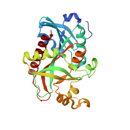
1RXS

E. coli uridine phosphorylase: 2'-deoxyuridine phosphate complex


Experimental Data Snapshot

  • Method: X-RAY DIFFRACTION
  • Resolution: 2.80 Å
  • R-Value Free: 0.218 
  • R-Value Work: 0.176 
  • R-Value Observed: 0.178 

wwPDB Validation   3D Report Full Report


This is version 1.4 of the entry. See complete history


Literature

Crystal structures of escherichia coli uridine phosphorylase in two native and three complexed forms reveal basis of substrate specificity, induced conformational changes and influence of potassium

Caradoc-Davies, T.T.Cutfield, S.M.Lamont, I.L.Cutfield, J.F.

(2004) J Mol Biol 337: 337-354

  • DOI: https://doi.org/10.1016/j.jmb.2004.01.039
  • Primary Citation of Related Structures:  
    1RXC, 1RXS, 1RXU, 1RXY, 1T0U

  • PubMed Abstract: 

    Uridine phosphorylase (UP) is a key enzyme in the pyrimidine salvage pathway that catalyses the reversible phosphorolysis of uridine to uracil and ribose 1-phosphate. Inhibiting liver UP in humans raises blood uridine levels and produces a protective effect ("uridine rescue") against the toxicity of the chemotherapeutic agent 5-fluorouracil without reducing its antitumour activity. We have investigated UP-substrate interactions by determining the crystal structures of native Escherichia coli UP (two forms), and complexes with 5-fluorouracil/ribose 1-phosphate, 2-deoxyuridine/phosphate and thymidine/phosphate. These hexameric structures confirm the overall structural similarity of UP to E.coli purine nucleoside phosphorylase (PNP) whereby, in the presence of substrate, each displays a closed conformation resulting from a concerted movement that closes the active site cleft. However, in contrast to PNP where helix segmentation is the major conformational change between the open and closed forms, in UP more extensive changes are observed. In particular a swinging movement of a flap region consisting of residues 224-234 seals the active site. This overall change in conformation results in compression of the active site cleft. Gln166 and Arg168, part of an inserted segment not seen in PNP, are key residues in the uracil binding pocket and together with a tightly bound water molecule are seen to be involved in the substrate specificity of UP. Enzyme activity shows a twofold dependence on potassium ion concentration. The presence of a potassium ion at the monomer/monomer interface induces some local rearrangement, which results in dimer stabilisation. The conservation of key residues and interactions with substrate in the phosphate and ribose binding pockets suggest that ribooxocarbenium ion formation during catalysis of UP may be similar to that proposed for E.coli PNP.


  • Organizational Affiliation

    Department of Biochemistry, Otago School of Medical Sciences, University of Otago, P.O. Box 56, Dunedin 9001, New Zealand. tom.caradoc-davies@stonebow.otago.ac.nz


Macromolecules
Find similar proteins by:  (by identity cutoff)  |  3D Structure
Entity ID: 1
MoleculeChains Sequence LengthOrganismDetailsImage
Uridine phosphorylase253Escherichia coliMutation(s): 0 
Gene Names: UDP
EC: 2.4.2.3
UniProt
Find proteins for P12758 (Escherichia coli (strain K12))
Explore P12758 
Go to UniProtKB:  P12758
Entity Groups  
Sequence Clusters30% Identity50% Identity70% Identity90% Identity95% Identity100% Identity
UniProt GroupP12758
Sequence Annotations
Expand
  • Reference Sequence
Small Molecules
Ligands 4 Unique
IDChains Name / Formula / InChI Key2D Diagram3D Interactions
V7O
Query on V7O

Download Ideal Coordinates CCD File 
AC [auth I]
AD [auth M]
EC [auth i]
FA [auth A]
FB [auth e]
AC [auth I],
AD [auth M],
EC [auth i],
FA [auth A],
FB [auth e],
GD [auth Q],
IC [auth J],
KD [auth o],
LA [auth B],
LB [auth R],
LC [auth j],
OA [auth b],
RC [auth k],
TB [auth H],
TC [auth L],
UA [auth c],
XB [auth h],
YA [auth D],
ZA [auth D],
ZC [auth M]
META VANADATE
O19 V7
CTUXFMMCMPZRLK-UHFFFAOYSA-N
DUR
Query on DUR

Download Ideal Coordinates CCD File 
CB [auth E]
DC [auth i]
DD [auth N]
EB [auth e]
FD [auth Q]
CB [auth E],
DC [auth i],
DD [auth N],
EB [auth e],
FD [auth Q],
HA [auth a],
HB [auth F],
HC [auth J],
ID [auth O],
KA [auth B],
KB [auth R],
KC [auth j],
NA [auth b],
OB [auth G],
OC [auth K],
QB [auth P],
QC [auth k],
RA [auth C],
SB [auth H],
TA [auth c],
VC [auth l],
WB [auth h],
XA [auth D],
YC [auth M],
ZB [auth I]
2'-DEOXYURIDINE
C9 H12 N2 O5
MXHRCPNRJAMMIM-SHYZEUOFSA-N
PO4
Query on PO4

Download Ideal Coordinates CCD File 
AB [auth E]
BC [auth i]
BD [auth N]
DB [auth e]
ED [auth Q]
AB [auth E],
BC [auth i],
BD [auth N],
DB [auth e],
ED [auth Q],
FC [auth J],
GA [auth a],
GB [auth F],
HD [auth O],
IA [auth B],
IB [auth R],
JC [auth j],
MA [auth b],
MB [auth G],
MC [auth K],
PA [auth C],
PB [auth P],
PC [auth k],
RB [auth H],
SA [auth c],
UB [auth h],
UC [auth l],
VA [auth D],
WC [auth M],
YB [auth I]
PHOSPHATE ION
O4 P
NBIIXXVUZAFLBC-UHFFFAOYSA-K
K
Query on K

Download Ideal Coordinates CCD File 
BB [auth E]
CC [auth i]
CD [auth N]
EA [auth A]
GC [auth J]
BB [auth E],
CC [auth i],
CD [auth N],
EA [auth A],
GC [auth J],
JA [auth B],
JB [auth R],
JD [auth o],
NB [auth G],
NC [auth K],
QA [auth C],
SC [auth L],
VB [auth h],
WA [auth D],
XC [auth M]
POTASSIUM ION
K
NPYPAHLBTDXSSS-UHFFFAOYSA-N
Experimental Data & Validation

Experimental Data

  • Method: X-RAY DIFFRACTION
  • Resolution: 2.80 Å
  • R-Value Free: 0.218 
  • R-Value Work: 0.176 
  • R-Value Observed: 0.178 
  • Space Group: P 1 21 1
Unit Cell:
Length ( Å )Angle ( ˚ )
a = 160.958α = 90
b = 98.212β = 109.09
c = 242.729γ = 90
Software Package:
Software NamePurpose
MOSFLMdata reduction
SCALAdata scaling
AMoREphasing
REFMACrefinement
CCP4data scaling

Structure Validation

View Full Validation Report


Entry History 

Deposition Data

Revision History  (Full details and data files)

  • Version 1.0: 2004-04-13
    Type: Initial release
  • Version 1.1: 2008-04-29
    Changes: Version format compliance
  • Version 1.2: 2011-07-13
    Changes: Advisory, Refinement description, Version format compliance
  • Version 1.3: 2024-03-13
    Changes: Advisory, Data collection, Database references, Derived calculations, Refinement description
  • Version 1.4: 2024-04-03
    Changes: Refinement description