1RV6

Crystal Structure of PlGF in Complex with Domain 2 of VEGFR1


Experimental Data Snapshot

  • Method: X-RAY DIFFRACTION
  • Resolution: 2.45 Å
  • R-Value Free: 0.260 
  • R-Value Work: 0.194 
  • R-Value Observed: 0.198 

Starting Model: experimental
View more details

wwPDB Validation   3D Report Full Report


This is version 1.3 of the entry. See complete history


Literature

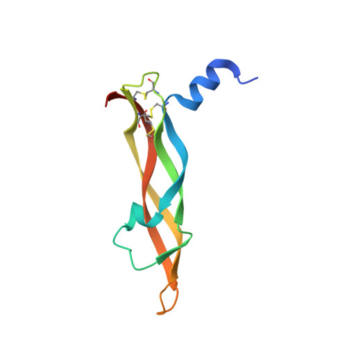
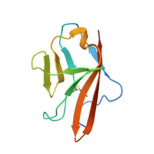
The crystal structure of placental growth factor in complex with domain 2 of vascular endothelial growth factor receptor-1.

Christinger, H.W.Fuh, G.de Vos, A.M.Wiesmann, C.

(2004) J Biol Chem 279: 10382-10388

  • DOI: https://doi.org/10.1074/jbc.M313237200
  • Primary Citation of Related Structures:  
    1RV6

  • PubMed Abstract: 

    Placental growth factor (PlGF) is a member of the vascular endothelial growth factor (VEGF) family and plays an important role in pathological angiogenic events. PlGF exerts its biological activities through binding to VEGFR1, a receptor tyrosine kinase that consists of seven immunoglobulin-like domains in its extracellular portion. Here we report the crystal structure of PlGF bound to the second immunoglobulin-like domain of VEGFR1 at 2.5 A resolution and compare the complex to the closely related structure of VEGF bound to the same receptor domain. The two growth factors, PlGF and VEGF, share a sequence identity of approximately 50%. Despite this moderate sequence conservation, they bind to the same binding interface of VEGFR1 in a very similar fashion, suggesting that both growth factors could induce very similar if not identical signaling events.


  • Organizational Affiliation

    Department of Protein Engineering Genentech, Inc., South San Francisco, California 94080, USA.


Macromolecules
Find similar proteins by:  (by identity cutoff)  |  3D Structure
Entity ID: 1
MoleculeChains Sequence LengthOrganismDetailsImage
placenta growth factor (PlGF)A [auth V],
B [auth W]
101Homo sapiensMutation(s): 0 
UniProt & NIH Common Fund Data Resources
Find proteins for P49763 (Homo sapiens)
Explore P49763 
Go to UniProtKB:  P49763
GTEx:  ENSG00000119630 
Entity Groups  
Sequence Clusters30% Identity50% Identity70% Identity90% Identity95% Identity100% Identity
UniProt GroupP49763
Sequence Annotations
Expand
  • Reference Sequence
Find similar proteins by:  (by identity cutoff)  |  3D Structure
Entity ID: 2
MoleculeChains Sequence LengthOrganismDetailsImage
FLT1 proteinC [auth X],
D [auth Y]
100Homo sapiensMutation(s): 0 
UniProt & NIH Common Fund Data Resources
Find proteins for P17948 (Homo sapiens)
Explore P17948 
Go to UniProtKB:  P17948
GTEx:  ENSG00000102755 
Entity Groups  
Sequence Clusters30% Identity50% Identity70% Identity90% Identity95% Identity100% Identity
UniProt GroupP17948
Sequence Annotations
Expand
  • Reference Sequence
Small Molecules
Ligands 1 Unique
IDChains Name / Formula / InChI Key2D Diagram3D Interactions
B3P
Query on B3P

Download Ideal Coordinates CCD File 
E [auth W]2-[3-(2-HYDROXY-1,1-DIHYDROXYMETHYL-ETHYLAMINO)-PROPYLAMINO]-2-HYDROXYMETHYL-PROPANE-1,3-DIOL
C11 H26 N2 O6
HHKZCCWKTZRCCL-UHFFFAOYSA-N
Experimental Data & Validation

Experimental Data

  • Method: X-RAY DIFFRACTION
  • Resolution: 2.45 Å
  • R-Value Free: 0.260 
  • R-Value Work: 0.194 
  • R-Value Observed: 0.198 
  • Space Group: P 21 21 21
Unit Cell:
Length ( Å )Angle ( ˚ )
a = 54.281α = 90
b = 71.979β = 90
c = 115.346γ = 90
Software Package:
Software NamePurpose
REFMACrefinement
DENZOdata reduction
SCALEPACKdata scaling
AMoREphasing

Structure Validation

View Full Validation Report


Entry History 

Deposition Data

Revision History  (Full details and data files)

  • Version 1.0: 2004-01-20
    Type: Initial release
  • Version 1.1: 2008-04-29
    Changes: Version format compliance
  • Version 1.2: 2011-07-13
    Changes: Advisory, Version format compliance
  • Version 1.3: 2023-08-23
    Changes: Data collection, Database references, Derived calculations, Refinement description