1RQ7

MYCOBACTERIUM TUBERCULOSIS FTSZ IN COMPLEX WITH GDP


Experimental Data Snapshot

  • Method: X-RAY DIFFRACTION
  • Resolution: 2.60 Å
  • R-Value Free: 0.242 
  • R-Value Work: 0.184 
  • R-Value Observed: 0.187 

wwPDB Validation   3D Report Full Report


Ligand Structure Quality Assessment 


This is version 1.5 of the entry. See complete history


Literature

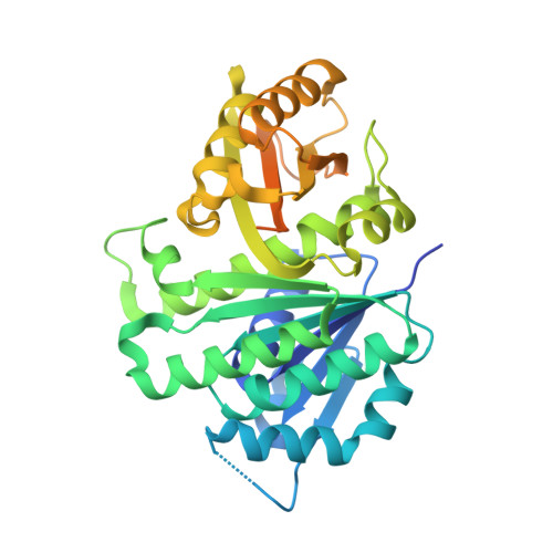
Structure of Mycobacterium tuberculosis FtsZ reveals unexpected, G protein-like conformational switches.

Leung, A.K.Lucile White, E.Ross, L.J.Reynolds, R.C.DeVito, J.A.Borhani, D.W.

(2004) J Mol Biol 342: 953-970

  • DOI: https://doi.org/10.1016/j.jmb.2004.07.061
  • Primary Citation of Related Structures:  
    1RLU, 1RQ2, 1RQ7

  • PubMed Abstract: 

    We report three crystal structures of the Mycobacterium tuberculosis cell division protein FtsZ, as the citrate, GDP, and GTPgammaS complexes, determined at 1.89, 2.60, and 2.08A resolution. MtbFtsZ crystallized as a tight, laterally oriented dimer distinct from the longitudinal polymer observed for alphabeta-tubulin. Mutational data on Escherichia coli FtsZ suggest that this dimer interface is important for proper protofilament and "Z-ring" assembly and function. An alpha-to-beta secondary structure conformational switch at the dimer interface is spatially analogous to, and has many of the hallmarks of, the Switch I conformational changes exhibited by G-proteins upon activation. The presence of a gamma-phosphate in the FtsZ active site modulates the conformation of the "tubulin" loop T3 (spatially analogous to the G-protein Switch II); T3 switching upon gamma-phosphate ligation is directly coupled to the alpha-to-beta switch by steric overlap. The dual conformational switches observed here for the first time in an FtsZ link GTP binding and hydrolysis to FtsZ (and tubulin) lateral assembly and Z-ring contraction, and they are suggestive of an underappreciated functional analogy between FtsZ, tubulin and G-proteins.


  • Organizational Affiliation

    Drug Discovery Division, Southern Research Institute, Birmingham, AL 35205, USA.


Macromolecules
Find similar proteins by:  (by identity cutoff)  |  3D Structure
Entity ID: 1
MoleculeChains Sequence LengthOrganismDetailsImage
Cell division protein ftsZ
A, B
382Mycobacterium tuberculosisMutation(s): 0 
Gene Names: FTSZRV2150CMT2209MTCY270.18MB2174C
UniProt
Find proteins for P9WN95 (Mycobacterium tuberculosis (strain ATCC 25618 / H37Rv))
Explore P9WN95 
Go to UniProtKB:  P9WN95
Entity Groups  
Sequence Clusters30% Identity50% Identity70% Identity90% Identity95% Identity100% Identity
UniProt GroupP9WN95
Sequence Annotations
Expand
  • Reference Sequence
Small Molecules
Ligands 1 Unique
IDChains Name / Formula / InChI Key2D Diagram3D Interactions
GDP
Query on GDP

Download Ideal Coordinates CCD File 
C [auth A]GUANOSINE-5'-DIPHOSPHATE
C10 H15 N5 O11 P2
QGWNDRXFNXRZMB-UUOKFMHZSA-N
Experimental Data & Validation

Experimental Data

  • Method: X-RAY DIFFRACTION
  • Resolution: 2.60 Å
  • R-Value Free: 0.242 
  • R-Value Work: 0.184 
  • R-Value Observed: 0.187 
  • Space Group: P 65
Unit Cell:
Length ( Å )Angle ( ˚ )
a = 88.619α = 90
b = 88.619β = 90
c = 179.196γ = 120
Software Package:
Software NamePurpose
REFMACrefinement
SCALAdata scaling
TRUNCATEdata reduction
X-PLORmodel building
X-PLORrefinement
CCP4data scaling
TRUNCATEdata scaling
X-PLORphasing
MOSFLMdata reduction

Structure Validation

View Full Validation Report


Ligand Structure Quality Assessment 


Entry History 

Deposition Data

Revision History  (Full details and data files)

  • Version 1.0: 2004-08-31
    Type: Initial release
  • Version 1.1: 2008-04-29
    Changes: Version format compliance
  • Version 1.2: 2011-07-13
    Changes: Version format compliance
  • Version 1.3: 2017-10-11
    Changes: Refinement description
  • Version 1.4: 2019-07-24
    Changes: Data collection, Refinement description
  • Version 1.5: 2023-08-23
    Changes: Data collection, Database references, Derived calculations, Refinement description