1ROM | pdb_00001rom

CRYSTAL STRUCTURE OF NITRIC REDUCTASE FROM DENITRIFYING FUNGUS FUSARIUM OXYSPORUM


Experimental Data Snapshot

  • Method: X-RAY DIFFRACTION
  • Resolution: 2.00 Å
  • R-Value Free: 
    0.266 (Depositor) 
  • R-Value Work: 
    0.197 (Depositor) 
  • R-Value Observed: 
    0.197 (Depositor) 

wwPDB Validation 3D Report Full Report

Validation slider image for 1ROM

This is version 1.3 of the entry. See complete history

Literature

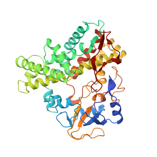
Crystal structure of nitric oxide reductase from denitrifying fungus Fusarium oxysporum.

Park, S.Y.Shimizu, H.Adachi, S.Nakagawa, A.Tanaka, I.Nakahara, K.Shoun, H.Obayashi, E.Nakamura, H.Iizuka, T.Shiro, Y.

(1997) Nat Struct Biol 4: 827-832

  • DOI: https://doi.org/10.1038/nsb1097-827
  • Primary Citation Related Structures: 
    1ROM, 2ROM

  • PubMed Abstract: 

    Structures of nitric oxide reductase (NOR) in the ferric resting and the ferrous CO states have been solved at 2.0 A resolution. These structures provide significant new insights into how NO is reduced in biological systems. The haem distal pocket is open to solvent, implicating this region as a possible NADH binding site. In combination with mutagenesis results, a hydrogen-bonding network from the water molecule adjacent to the iron ligand to the protein surface of the distal pocket through the hydroxyl group of Ser 286 and the carboxyl group of Asp 393 can be assigned to a pathway for proton delivery during the NO reduction reaction.


  • Organizational Affiliation
    • Institute of Physical and Chemical Research (RIKEN), Saitama, Japan.

Macromolecule Content 

  • Total Structure Weight: 45.04 kDa 
  • Atom Count: 3,301 
  • Modeled Residue Count: 399 
  • Deposited Residue Count: 403 
  • Unique protein chains: 1

Macromolecules

Find similar proteins by:|  3D Structure
Entity ID: 1
MoleculeChains  Sequence LengthOrganismDetailsImage
CYTOCHROME P450403Fusarium oxysporumMutation(s): 0 
EC: 1.14 (PDB Primary Data), 1.7.1.14 (UniProt)
UniProt
Find proteins for P23295 (Fusarium oxysporum)
Explore P23295 
Go to UniProtKB:  P23295
Entity Groups
Sequence Clusters30% Identity50% Identity70% Identity90% Identity95% Identity100% Identity
UniProt GroupP23295
Sequence Annotations
Expand
Reference Sequence

Small Molecules

Ligands 1 Unique
IDChains Name / Formula / InChI Key2D Diagram3D Interactions
HEM

Query on HEM


Download:Ideal Coordinates CCD File
B [auth A]PROTOPORPHYRIN IX CONTAINING FE
C34 H32 Fe N4 O4
KABFMIBPWCXCRK-RGGAHWMASA-L

Experimental Data & Validation

Experimental Data

  • Method: X-RAY DIFFRACTION
  • Resolution: 2.00 Å
  • R-Value Free:  0.266 (Depositor) 
  • R-Value Work:  0.197 (Depositor) 
  • R-Value Observed: 0.197 (Depositor) 
Space Group: P 21 21 21
Unit Cell:
Length ( Å )Angle ( ˚ )
a = 54.99α = 90
b = 82.66β = 90
c = 87.21γ = 90
Software Package:
Software NamePurpose
X-PLORmodel building
X-PLORrefinement
DENZOdata reduction
CCP4data scaling
Agrovatadata scaling
X-PLORphasing

Structure Validation

View Full Validation Report


Entry History 

Deposition Data

Revision History  (Full details and data files)

  • Version 1.0: 1997-10-15
    Type: Initial release
  • Version 1.1: 2008-03-24
    Changes: Version format compliance
  • Version 1.2: 2011-07-13
    Changes: Version format compliance
  • Version 1.3: 2024-02-14
    Changes: Data collection, Database references, Derived calculations, Other, Refinement description