1ROB | pdb_00001rob

STRUCTURE OF THE CRYSTALLINE COMPLEX OF CYTIDYLIC ACID (2'-CMP) WITH RIBONUCLEASE AT 1.6 ANGSTROMS RESOLUTION


Experimental Data Snapshot

  • Method: X-RAY DIFFRACTION
  • Resolution: 1.60 Å
  • R-Value Work: 
    0.237 (DCC) 
  • R-Value Observed: 
    0.170 (Depositor) 

wwPDB Validation 3D Report Full Report

Validation slider image for 1ROB

Ligand Structure Quality Assessment 


This is version 1.4 of the entry. See complete history

Literature

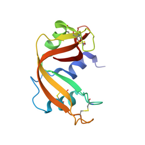
Structure of the crystalline complex of cytidylic acid (2'-CMP) with ribonuclease at 1.6 A resolution. Conservation of solvent sites in RNase-A high-resolution structures.

Lisgarten, J.N.Gupta, V.Maes, D.Wyns, L.Zegers, I.Palmer, R.A.Dealwis, C.G.Aguilar, C.F.Hemmings, A.M.

(1993) Acta Crystallogr D Biol Crystallogr 49: 541-547

  • DOI: https://doi.org/10.1107/S090744499300719X
  • Primary Citation Related Structures: 
    1ROB

  • PubMed Abstract: 

    The X-ray structure of the inhibitor complex of bovine ribonuclease A with cytidylic acid (2'-CMP) has been determined at 1.6 A resolution and refined by restrained least squares to R = 0.17 for 11 945 reflections. Binding of the inhibitor molecule to the protein is confirmed to be in the productive mode associated with enzyme activity. A study of conserved solvent sites amongst high-resolution structures in the same crystal form reveals a stabilizing water cluster between the N and C termini.


  • Organizational Affiliation
    • Department of Ultrastructure, Institut voor Molekulaire Biologie, Vrije Universiteit Brussel, Sint Genesius Rode, Belgium.

Macromolecule Content 

  • Total Structure Weight: 14.03 kDa 
  • Atom Count: 1,077 
  • Modeled Residue Count: 124 
  • Deposited Residue Count: 124 
  • Unique protein chains: 1

Macromolecules

Find similar proteins by:|  3D Structure
Entity ID: 1
MoleculeChains  Sequence LengthOrganismDetailsImage
RIBONUCLEASE A124Bos taurusMutation(s): 0 
EC: 3.1.27.5 (PDB Primary Data), 4.6.1.18 (UniProt)
UniProt
Find proteins for P61823 (Bos taurus)
Explore P61823 
Go to UniProtKB:  P61823
Entity Groups
Sequence Clusters30% Identity50% Identity70% Identity90% Identity95% Identity100% Identity
UniProt GroupP61823
Sequence Annotations
Expand
Reference Sequence

Small Molecules

Ligands 1 Unique
IDChains Name / Formula / InChI Key2D Diagram3D Interactions
C2P

Query on C2P


Download:Ideal Coordinates CCD File
B [auth A]CYTIDINE-2'-MONOPHOSPHATE
C9 H14 N3 O8 P
YQUAKORMLHPSLZ-XVFCMESISA-N

Experimental Data & Validation

Experimental Data

  • Method: X-RAY DIFFRACTION
  • Resolution: 1.60 Å
  • R-Value Work:  0.237 (DCC) 
  • R-Value Observed: 0.170 (Depositor) 
Space Group: P 1 21 1
Unit Cell:
Length ( Å )Angle ( ˚ )
a = 30.512α = 90
b = 38.242β = 106.1
c = 53.303γ = 90
Software Package:
Software NamePurpose
RESTRAINrefinement

Structure Validation

View Full Validation Report


Ligand Structure Quality Assessment 


Entry History 

Deposition Data

Revision History  (Full details and data files)

  • Version 1.0: 1994-01-31
    Type: Initial release
  • Version 1.1: 2008-03-24
    Changes: Version format compliance
  • Version 1.2: 2011-07-13
    Changes: Version format compliance
  • Version 1.3: 2024-06-05
    Changes: Data collection, Database references, Derived calculations, Other
  • Version 1.4: 2024-10-23
    Changes: Structure summary