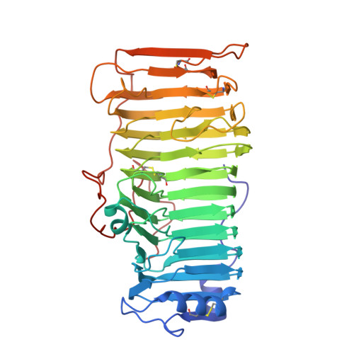
1RMG

RHAMNOGALACTURONASE A FROM ASPERGILLUS ACULEATUS


Experimental Data Snapshot

  • Method: X-RAY DIFFRACTION
  • Resolution: 2.00 Å
  • R-Value Free: 0.214 
  • R-Value Work: 0.174 
  • R-Value Observed: 0.174 

wwPDB Validation   3D Report Full Report


Ligand Structure Quality Assessment 


This is version 2.0 of the entry. See complete history


Literature

The crystal structure of rhamnogalacturonase A from Aspergillus aculeatus: a right-handed parallel beta helix.

Petersen, T.N.Kauppinen, S.Larsen, S.

(1997) Structure 5: 533-544

  • DOI: https://doi.org/10.1016/s0969-2126(97)00209-8
  • Primary Citation of Related Structures:  
    1RMG

  • PubMed Abstract: 

    Pectic substances are the major polysaccharide components of the middle lamella and primary cell wall of dicotyledonous plants. They consist of homogalacturonan 'smooth' regions and highly rhamnified 'hairy' regions of rhamnogalacturonan. The backbone in rhamnogalacturonan-l (RG-l), which is composed of alternating galacturonic acid and rhamnose residues, is the substrate for a new class of enzymes known as rhamnogalacturnoases (RGases). RGase A is a novel enzyme implicated in the enzymatic degradation of RG-l. The structure of RGase A from Aspergillus aculeatus has been solved by the single isomorphous replacement method including anomalous scattering (SIRAS method) to 2.0 A resolution. The enzyme folds into a large right-handed parallel beta helix, with a core composed of 13 turns of beta strands. Four parallel beta sheets (PB1, PB1a, PB2 and PB3), formed by the consecutive turns, are typically separated by a residue in the conformation of a left-handed alpha helix. As a consequence of the consecutive turns, 32% of all residues have their sidechains aligned at the surface or in the interior of the parallel beta helix. The aligned residues at the surface are dominated by threonine, aspartic acid and asparagine, whereas valine, leucine and isoleucine are most frequently found in the interior. A very large hydrophobic cavity is found in the interior of the parallel beta helix. The potential active site is a groove, oriented almost perpendicular to the helical axis, containing a cluster of three aspartic acid residues and one glutamic acid residue. The enzyme is highly glycosylated; two N-linked and eighteen O-linked glycosylation sites have been found in the structure. Rhamnogalacturonase A from A. aculeatus is the first three-dimensional structure of an enzyme hydrolyzing glycoside bonds within the backbone of RG-l. The large groove, which is the potential active site of RGase A, is also seen in the structures of pectate lyases. Two catalytic aspartic acid residues, which have been proposed to have a catalytic role, reside in this area of RGase A. The distance between the aspartic acid residues is consistent with the inverting mechanism of catalysis. The glycan groups bound to RGase A are important to the stability of the crystal, as the carbohydrate moiety is involved in most of the intermolecular hydrogen bonds.


  • Organizational Affiliation

    Centre for Crystallographic Studies, Department of Chemistry, University of Copenhagen, Universitetsparken 5, 2100, Copenhagen, Denmark.


Macromolecules
Find similar proteins by:  (by identity cutoff)  |  3D Structure
Entity ID: 1
MoleculeChains Sequence LengthOrganismDetailsImage
RHAMNOGALACTURONASE A422Aspergillus aculeatusMutation(s): 0 
EC: 3.2.1
UniProt
Find proteins for Q00001 (Aspergillus aculeatus)
Explore Q00001 
Go to UniProtKB:  Q00001
Entity Groups  
Sequence Clusters30% Identity50% Identity70% Identity90% Identity95% Identity100% Identity
UniProt GroupQ00001
Glycosylation
Glycosylation Sites: 17
Sequence Annotations
Expand
  • Reference Sequence
Oligosaccharides

Help

Entity ID: 2
MoleculeChains Length2D Diagram Glycosylation3D Interactions
alpha-D-mannopyranose-(1-3)-[beta-D-mannopyranose-(1-6)]beta-D-mannopyranose-(1-4)-2-acetamido-2-deoxy-beta-D-glucopyranose-(1-4)-2-acetamido-2-deoxy-beta-D-glucopyranose
B
5N-Glycosylation
Glycosylation Resources
GlyTouCan:  G51945PF
GlyCosmos:  G51945PF
GlyGen:  G51945PF
Entity ID: 3
MoleculeChains Length2D Diagram Glycosylation3D Interactions
beta-D-mannopyranose-(1-4)-2-acetamido-2-deoxy-beta-D-glucopyranose-(1-4)-2-acetamido-2-deoxy-beta-D-glucopyranose
C
3N-Glycosylation
Glycosylation Resources
GlyTouCan:  G15407YE
GlyCosmos:  G15407YE
GlyGen:  G15407YE
Small Molecules
Ligands 3 Unique
IDChains Name / Formula / InChI Key2D Diagram3D Interactions
GLC
Query on GLC

Download Ideal Coordinates CCD File 
K [auth A]alpha-D-glucopyranose
C6 H12 O6
WQZGKKKJIJFFOK-DVKNGEFBSA-N
BMA
Query on BMA

Download Ideal Coordinates CCD File 
E [auth A],
M [auth A],
Q [auth A]
beta-D-mannopyranose
C6 H12 O6
WQZGKKKJIJFFOK-RWOPYEJCSA-N
MAN
Query on MAN

Download Ideal Coordinates CCD File 
D [auth A]
F [auth A]
G [auth A]
H [auth A]
I [auth A]
D [auth A],
F [auth A],
G [auth A],
H [auth A],
I [auth A],
J [auth A],
L [auth A],
N [auth A],
O [auth A],
P [auth A],
R [auth A],
S [auth A],
T [auth A],
U [auth A]
alpha-D-mannopyranose
C6 H12 O6
WQZGKKKJIJFFOK-PQMKYFCFSA-N
Experimental Data & Validation

Experimental Data

  • Method: X-RAY DIFFRACTION
  • Resolution: 2.00 Å
  • R-Value Free: 0.214 
  • R-Value Work: 0.174 
  • R-Value Observed: 0.174 
  • Space Group: I 2 2 2
Unit Cell:
Length ( Å )Angle ( ˚ )
a = 62.9α = 90
b = 125.4β = 90
c = 137γ = 90
Software Package:
Software NamePurpose
DENZOdata reduction
CCP4-PROGRAMSdata reduction
X-PLORmodel building
X-PLORrefinement
CCP4data scaling
X-PLORphasing

Structure Validation

View Full Validation Report


Ligand Structure Quality Assessment 


Entry History 

Deposition Data

Revision History  (Full details and data files)

  • Version 1.0: 1998-03-04
    Type: Initial release
  • Version 1.1: 2008-03-24
    Changes: Version format compliance
  • Version 1.2: 2011-07-13
    Changes: Non-polymer description, Version format compliance
  • Version 2.0: 2020-07-29
    Type: Remediation
    Reason: Carbohydrate remediation
    Changes: Atomic model, Data collection, Derived calculations, Structure summary