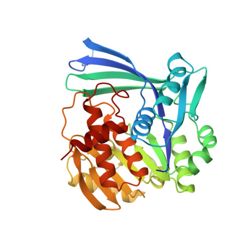
Structure of Escherichia coli ribokinase in complex with ribose and dinucleotide determined to 1.8 A resolution: insights into a new family of kinase structures.
Sigrell, J.A., Cameron, A.D., Jones, T.A., Mowbray, S.L.(1998) Structure 6: 183-193
- PubMed: 9519409 Search on PubMed
- DOI: https://doi.org/10.1016/s0969-2126(98)00020-3
- Primary Citation Related Structures:
1RKD - PubMed Abstract:
D-ribose must be phosphorylated at O5' before it can be used in either anabolism or catabolism. This reaction is catalysed by ribokinase and requires the presence of ATP and magnesium. Ribokinase is a member of a family of carbohydrate kinases of previously unknown structure. The crystal structure of ribokinase from Escherichia coli in complex with ribose and dinucleotide was determined at 1.84 A resolution by multiple isomorphous replacement. There is one 33 kDa monomer of ribokinase in the asymmetric unit but the protein forms a dimer around a crystallographic twofold axis. Each subunit consists of a central alpha/beta unit, with a new type of nucleotide-binding fold, and a distinct beta sheet that forms a lid over the ribose-binding site. Contact between subunits involves orthogonal packing of beta sheets, in a novel dimer interaction that we call a beta clasp. Inspection of the complex indicates that ribokinase utilises both a catalytic base for activation of the ribose in nucleophilic attack and an anion hole that stabilises the transition state during phosphoryl transfer. The structure suggests an ordered reaction mechanism, similar to those proposed for other carbohydrate kinases that probably involves conformational changes. We propose that the beta-clasp structure acts as a lid, closing and opening upon binding and release of ribose. From these observations, an understanding of the structure and catalytic mechanism of related sugar kinases can be obtained.
- Department of Molecular Biology, Uppsala University, Sweden.
Organizational Affiliation: