Paramagnetism-based refinement strategy for the solution structure of human alpha-parvalbumin
Baig, I., Bertini, I., Del Bianco, C., Gupta, Y.K., Lee, Y.-M., Luchinat, C., Quattrone, A.(2004) Biochemistry 43: 5562-5573
- PubMed: 15122922 Search on PubMed
- DOI: https://doi.org/10.1021/bi035879k
- Primary Citation Related Structures:
1RJV, 1RK9 - PubMed Abstract:
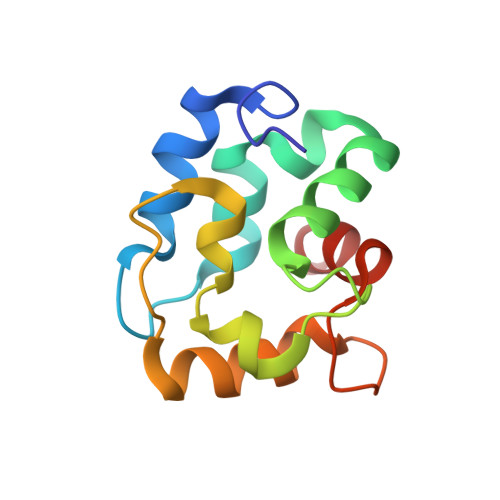
In the frame of a research aimed at the detailed structural characterization of human calcium-binding proteins of the EF-hand family, the solution structure of human alpha-parvalbumin has been solved by NMR and refined with the help of substitution of the Ca(2+) ion in the EF site with the paramagnetic Dy(3+) ion. A simple (1)H-(15)N HSQC spectrum allowed the NH assignments based on the properties of Dy(3+). This allowed us to exploit pseudocontact shifts and residual dipolar couplings for solution structure refinement. The backbone and heavy atom RMSD are 0.55 +/- 0.08 and 1.02 +/- 0.08 A, respectively, and decrease to 0.39 +/- 0.05 and 0.90 +/- 0.06 A upon refinement with paramagnetism-based restraints. The RMSD for the metal itself in the EF site in the refined structure is 0.26 +/- 0.12 A. Backbone NH R(1), R(2), and NOE measured at two temperatures show the protein to be relatively rigid. The NH orientations are well determined by the paramagnetism-based restraints. This allows us to detect small but significant local structural differences with the orthologue protein from rat, whose X-ray structure is available at 2.0 A resolution. All differences are related to local changes in the amino acidic composition.
- Magnetic Resonance Centre and Department of Chemistry, University of Florence, Via Luigi Sacconi 6, 50019 Sesto Fiorentino, Italy.
Organizational Affiliation: