The functional role of the binuclear metal center in D-aminoacylase: one-metal activation and second-metal attenuation.
Lai, W.L., Chou, L.Y., Ting, C.Y., Kirby, R., Tsai, Y.C., Wang, A.H., Liaw, S.H.(2004) J Biological Chem 279: 13962-13967
- PubMed: 14736882 Search on PubMed
- DOI: https://doi.org/10.1074/jbc.M308849200
- Primary Citation Related Structures:
1RJP, 1RJQ, 1RJR, 1RK5, 1RK6, 1V4Y, 1V51 - PubMed Abstract:
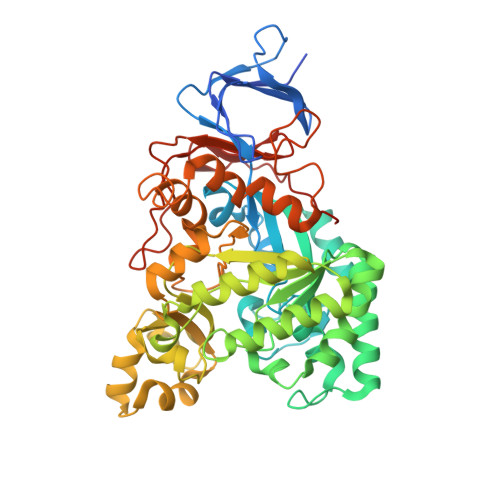
Our structural comparison of the TIM barrel metal-dependent hydrolase(-like) superfamily suggests a classification of their divergent active sites into four types: alphabeta-binuclear, alpha-mononuclear, beta-mononuclear, and metal-independent subsets. The d-aminoacylase from Alcaligenes faecalis DA1 belongs to the beta-mononuclear subset due to the fact that the catalytically essential Zn(2+) is tightly bound at the beta site with coordination by Cys(96), His(220), and His(250), even though it possesses a binuclear active site with a weak alpha binding site. Additional Zn(2+), Cd(2+), and Cu(2+), but not Ni(2+), Co(2+), Mg(2+), Mn(2+), and Ca(2+), can inhibit enzyme activity. Crystal structures of these metal derivatives show that Zn(2+) and Cd(2+) bind at the alpha(1) subsite ligated by His(67), His(69), and Asp(366), while Cu(2+) at the alpha(2) subsite is chelated by His(67), His(69) and Cys(96). Unexpectedly, the crystal structure of the inactive H220A mutant displays that the endogenous Zn(2+) shifts to the alpha(3) subsite coordinated by His(67), His(69), Cys(96), and Asp(366), revealing that elimination of the beta site changes the coordination geometry of the alpha ion with an enhanced affinity. Kinetic studies of the metal ligand mutants such as C96D indicate the uniqueness of the unusual bridging cysteine and its involvement in catalysis. Therefore, the two metal-binding sites in the d-aminoacylase are interactive with partially mutual exclusion, thus resulting in widely different affinities for the activation/attenuation mechanism, in which the enzyme is activated by the metal ion at the beta site, but inhibited by the subsequent binding of the second ion at the alpha site.
- Structural Biology Program, Institute of Biochemistry, Faculty of Life Science, National Yang-Ming University, Taipei 11221, Taiwan.
Organizational Affiliation: