1RHL | pdb_00001rhl

RIBONUCLEASE T1 COMPLEXED WITH 2'GMP/G23A MUTANT


Experimental Data Snapshot

  • Method: X-RAY DIFFRACTION
  • Resolution: 1.95 Å
  • R-Value Free: 
    0.208 (Depositor), 0.207 (DCC) 
  • R-Value Work: 
    0.164 (Depositor), 0.165 (DCC) 
  • R-Value Observed: 
    0.164 (Depositor) 

Starting Model: experimental
View more details

wwPDB Validation 3D Report Full Report

Validation slider image for 1RHL

Ligand Structure Quality Assessment 


This is version 1.5 of the entry. See complete history

Literature

Hydrogen-exchange stabilities of RNase T1 and variants with buried and solvent-exposed Ala --> Gly mutations in the helix.

Huyghues-Despointes, B.M.Langhorst, U.Steyaert, J.Pace, C.N.Scholtz, J.M.

(1999) Biochemistry 38: 16481-16490

  • DOI: https://doi.org/10.1021/bi9919450
  • Primary Citation Related Structures: 
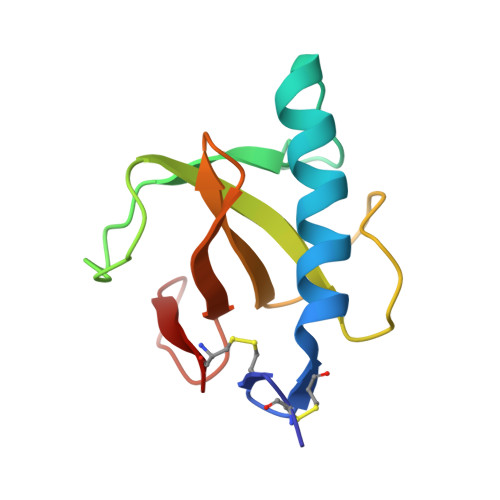
    1RHL

  • PubMed Abstract: 

    Hydrogen-exchange rates were measured for RNase T1 and three variants with Ala --> Gly substitutions at a solvent-exposed (residue 21) and a buried (residue 23) position in the helix: A21G, G23A, and A21G + G23A. These results were used to measure the stabilities of the proteins. The hydrogen-exchange stabilities (DeltaG(HX)) for the most stable residues in each variant agree with the equilibrium conformational stability measured by urea denaturation (DeltaG(U)), if the effects of D(2)O and proline isomerization are included [Huyghues-Despointes, B. M. P., Scholtz, J. M., and Pace, C. N. (1999) Nat. Struct. Biol. 6, 210-212]. These residues also show similar changes in DeltaG(HX) upon Ala --> Gly mutations (DeltaDeltaG(HX)) as compared to equilibrium measurements (DeltaDeltaG(U)), indicating that the most stable residues are exchanging from the globally unfolded ensemble. Alanine is stabilizing compared to glycine by 1 kcal/mol at a solvent-exposed site 21 as seen by other methods for the RNase T1 protein and peptide helix [Myers, J. K., Pace, C. N., and Scholtz, J. M. (1997) Proc. Natl. Acad. Sci. U.S.A. 94, 3833-2837], while it is destabilizing at the buried site 23 by the same amount. For the A21G variant, only local NMR chemical shift perturbations are observed compared to RNase T1. For the G23A variant, large chemical shift changes are seen throughout the sequence, although X-ray crystal structures of the variant and RNase T1 are nearly superimposable. Ala --> Gly mutations in the helix of RNase T1 at both helical positions alter the native-state hydrogen-exchange stabilities of residues throughout the sequence.


  • Organizational Affiliation
    • Department of Medical Biochemistry and Genetics, Center for Macromolecular Design, Texas A&M University, College Station 77843, USA.

Macromolecule Content 

  • Total Structure Weight: 11.51 kDa 
  • Atom Count: 886 
  • Modeled Residue Count: 104 
  • Deposited Residue Count: 104 
  • Unique protein chains: 1

Macromolecules

Find similar proteins by:|  3D Structure
Entity ID: 1
MoleculeChains  Sequence LengthOrganismDetailsImage
PROTEIN (RIBONUCLEASE T1)104Aspergillus oryzaeMutation(s): 1 
EC: 3.1.27.3 (PDB Primary Data), 4.6.1.24 (UniProt)
UniProt
Find proteins for P00651 (Aspergillus oryzae (strain ATCC 42149 / RIB 40))
Explore P00651 
Go to UniProtKB:  P00651
Entity Groups
Sequence Clusters30% Identity50% Identity70% Identity90% Identity95% Identity100% Identity
UniProt GroupP00651
Sequence Annotations
Expand
Reference Sequence

Small Molecules

Experimental Data & Validation

Experimental Data

  • Method: X-RAY DIFFRACTION
  • Resolution: 1.95 Å
  • R-Value Free:  0.208 (Depositor), 0.207 (DCC) 
  • R-Value Work:  0.164 (Depositor), 0.165 (DCC) 
  • R-Value Observed: 0.164 (Depositor) 
Space Group: P 21 21 21
Unit Cell:
Length ( Å )Angle ( ˚ )
a = 40.44α = 90
b = 48.23β = 90
c = 50.95γ = 90
Software Package:
Software NamePurpose
DENZOdata reduction
SCALEPACKdata scaling
X-PLORmodel building
X-PLORrefinement
X-PLORphasing

Structure Validation

View Full Validation Report


Ligand Structure Quality Assessment 


Entry History 

Deposition Data

Revision History  (Full details and data files)

  • Version 1.0: 1998-10-14
    Type: Initial release
  • Version 1.1: 2007-10-16
    Changes: Version format compliance
  • Version 1.2: 2011-07-13
    Changes: Version format compliance
  • Version 1.3: 2021-11-03
    Changes: Database references, Derived calculations
  • Version 1.4: 2023-08-23
    Changes: Data collection, Refinement description
  • Version 1.5: 2024-10-30
    Changes: Structure summary