Ribonuclease from Streptomyces aureofaciens at atomic resolution.
Sevcik, J., Dauter, Z., Lamzin, V.S., Wilson, K.S.(1996) Acta Crystallogr D Biol Crystallogr 52: 327-344
- PubMed: 15299705 Search on PubMed
- DOI: https://doi.org/10.1107/S0907444995007669
- Primary Citation Related Structures:
1RGE, 1RGF, 1RGG, 1RGH - PubMed Abstract:
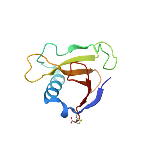
Crystals of ribonuclease from Streptomyces aureofaciens diffract to atomic resolution at room temperature. Using synchrotron radiation and an imaging-plate scanner, X-ray data have been recorded to 1.20 A resolution from a crystal of native enzyme and to 1.15 A from a crystal of a complex with guanosine-2'-monophosphate. Refinement with anisotropic atomic temperature factors resulted in increased accuracy of the structure. The R factors for the two structures are 10.6 and 10.9%. The estimated r.m.s. error in the coordinates is 0.05 A, less than half that obtained in the previous analysis at 1.7 A resolution. For the well ordered part of the main chain the error falls to below 0.02 A as estimated from inversion of the least-squares matrix. The two independent molecules in the asymmetric unit allowed detailed analysis of peptide planarity and some torsion angles. The high accuracy of the analysis revealed density for a partially occupied anion in the nucleotide binding site of molecule A in the native structure which was not seen at lower resolution. The anisotropic model allowed correction of the identity of the residue at position 72 from cysteine to threonine. Cys72 SG had been modelled in previous analyses with two conformations. The solvent structure was modelled by means of an automated procedure employing a set of objective criteria. The solvent structure for models refined using different programs with isotropic and anisotropic description of thermal motion is compared.
- Institute of Molecular Biology, Slovak Academy of Sciences, Bratislava, Slovak Republic.
Organizational Affiliation: