1RFN

HUMAN COAGULATION FACTOR IXA IN COMPLEX WITH P-AMINO BENZAMIDINE


Experimental Data Snapshot

  • Method: X-RAY DIFFRACTION
  • Resolution: 2.80 Å
  • R-Value Free: 0.273 
  • R-Value Work: 0.216 

wwPDB Validation   3D Report Full Report


This is version 1.3 of the entry. See complete history


Literature

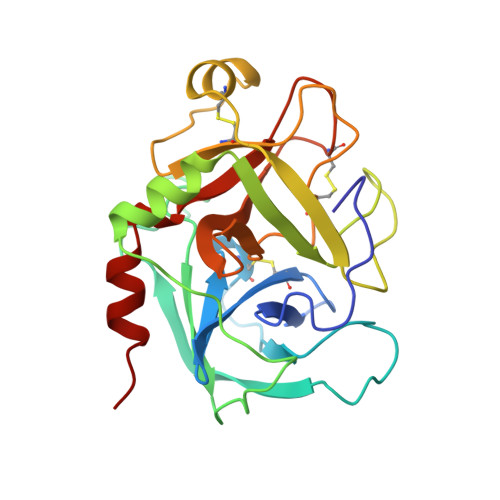
Coagulation factor IXa: the relaxed conformation of Tyr99 blocks substrate binding.

Hopfner, K.P.Lang, A.Karcher, A.Sichler, K.Kopetzki, E.Brandstetter, H.Huber, R.Bode, W.Engh, R.A.

(1999) Structure 7: 989-996

  • DOI: https://doi.org/10.1016/s0969-2126(99)80125-7
  • Primary Citation of Related Structures:  
    1RFN

  • PubMed Abstract: 

    Among the S1 family of serine proteinases, the blood coagulation factor IXa (fIXa) is uniquely inefficient against synthetic peptide substrates. Mutagenesis studies show that a loop of residues at the S2-S4 substrate-binding cleft (the 99-loop) contributes to the low efficiency. The crystal structure of porcine fIXa in complex with the inhibitor D-Phe-Pro-Arg-chloromethylketone (PPACK) was unable to directly clarify the role of the 99-loop, as the doubly covalent inhibitor induced an active conformation of fIXa. The crystal structure of a recombinant two-domain construct of human fIXa in complex with p-aminobenzamidine shows that the Tyr99 sidechain adopts an atypical conformation in the absence of substrate interactions. In this conformation, the hydroxyl group occupies the volume corresponding to the mainchain of a canonically bound substrate P2 residue. To accommodate substrate binding, Tyr99 must adopt a higher energy conformation that creates the S2 pocket and restricts the S4 pocket, as in fIXa-PPACK. The energy cost may contribute significantly to the poor K(M) values of fIXa for chromogenic substrates. In homologs, such as factor Xa and tissue plasminogen activator, the different conformation of the 99-loop leaves Tyr99 in low-energy conformations in both bound and unbound states. Molecular recognition of substrates by fIXa seems to be determined by the action of the 99-loop on Tyr99. This is in contrast to other coagulation enzymes where, in general, the chemical nature of residue 99 determines molecular recognition in S2 and S3-S4. This dominant role on substrate interaction suggests that the 99-loop may be rearranged in the physiological fX activation complex of fIXa, fVIIIa, and fX.


  • Organizational Affiliation

    Abteilung Strukturforschung, Max-Planck-Institut für Biochemie, Martinsried, Germany.


Macromolecules
Find similar proteins by:  (by identity cutoff)  |  3D Structure
Entity ID: 1
MoleculeChains Sequence LengthOrganismDetailsImage
PROTEIN (COAGULATION FACTOR IX)235Homo sapiensMutation(s): 0 
UniProt & NIH Common Fund Data Resources
Find proteins for P00740 (Homo sapiens)
Explore P00740 
Go to UniProtKB:  P00740
PHAROS:  P00740
GTEx:  ENSG00000101981 
Entity Groups  
Sequence Clusters30% Identity50% Identity70% Identity90% Identity95% Identity100% Identity
UniProt GroupP00740
Sequence Annotations
Expand
  • Reference Sequence
Find similar proteins by:  (by identity cutoff)  |  3D Structure
Entity ID: 2
MoleculeChains Sequence LengthOrganismDetailsImage
PROTEIN (COAGULATION FACTOR IX)57Homo sapiensMutation(s): 0 
UniProt & NIH Common Fund Data Resources
Find proteins for P00740 (Homo sapiens)
Explore P00740 
Go to UniProtKB:  P00740
PHAROS:  P00740
GTEx:  ENSG00000101981 
Entity Groups  
Sequence Clusters30% Identity50% Identity70% Identity90% Identity95% Identity100% Identity
UniProt GroupP00740
Sequence Annotations
Expand
  • Reference Sequence
Small Molecules
Ligands 3 Unique
IDChains Name / Formula / InChI Key2D Diagram3D Interactions
PBZ
Query on PBZ

Download Ideal Coordinates CCD File 
D [auth A]P-AMINO BENZAMIDINE
C7 H10 N3
WPANETAWYGDRLL-UHFFFAOYSA-O
TBU
Query on TBU

Download Ideal Coordinates CCD File 
E [auth A]TERTIARY-BUTYL ALCOHOL
C4 H10 O
DKGAVHZHDRPRBM-UHFFFAOYSA-N
CA
Query on CA

Download Ideal Coordinates CCD File 
C [auth A]CALCIUM ION
Ca
BHPQYMZQTOCNFJ-UHFFFAOYSA-N
Experimental Data & Validation

Experimental Data

  • Method: X-RAY DIFFRACTION
  • Resolution: 2.80 Å
  • R-Value Free: 0.273 
  • R-Value Work: 0.216 
  • Space Group: P 61 2 2
Unit Cell:
Length ( Å )Angle ( ˚ )
a = 90.9α = 90
b = 90.9β = 90
c = 151.6γ = 120
Software Package:
Software NamePurpose
X-PLORrefinement

Structure Validation

View Full Validation Report


Entry History 

Deposition Data

Revision History  (Full details and data files)

  • Version 1.0: 1999-09-01
    Type: Initial release
  • Version 1.1: 2008-04-26
    Changes: Version format compliance
  • Version 1.2: 2011-07-13
    Changes: Version format compliance
  • Version 1.3: 2023-12-27
    Changes: Data collection, Database references, Derived calculations