1RAC

CRYSTAL STRUCTURE OF CTP-LIGATED T STATE ASPARTATE TRANSCARBAMOYLASE AT 2.5 ANGSTROMS RESOLUTION: IMPLICATIONS FOR ATCASE MUTANTS AND THE MECHANISM OF NEGATIVE COOPERATIVITY


Experimental Data Snapshot

  • Method: X-RAY DIFFRACTION
  • Resolution: 2.50 Å
  • R-Value Work: 0.189 
  • R-Value Observed: 0.189 

wwPDB Validation   3D Report Full Report


This is version 1.5 of the entry. See complete history


Literature

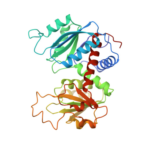
Crystal structure of CTP-ligated T state aspartate transcarbamoylase at 2.5 A resolution: implications for ATCase mutants and the mechanism of negative cooperativity.

Kosman, R.P.Gouaux, J.E.Lipscomb, W.N.

(1993) Proteins 15: 147-176

  • DOI: https://doi.org/10.1002/prot.340150206
  • Primary Citation of Related Structures:  
    1RAA, 1RAB, 1RAC, 1RAD, 1RAE, 1RAF, 1RAG, 1RAH, 1RAI

  • PubMed Abstract: 

    The X-ray crystal structure of CTP-ligated T state aspartate transcarbamoylase has been refined to an R factor of 0.182 at 2.5 A resolution using the computer program X-PLOR. The structure contains 81 sites for solvent and has rms deviations from ideality in bond lengths and bond angles of 0.018 A and 3.722 degrees, respectively. The cytosine base of CTP interacts with the main chain carbonyl oxygens of rTyr-89 and rIle-12, the main chain NH of rIle-12, and the amino group of rLys-60. The ribose hydroxyls form polar contacts with the amino group of rLys-60, a carboxylate oxygen of rAsp-19, and the main chain carbonyl oxygen of rVal-9. The phosphate oxygens of CTP interact with the amino group of rLys-94, the hydroxyl of rThr-82, and an imidazole nitrogen of rHis-20. Recent mutagenesis experiments evaluated in parallel with the structure reported here indicate that alterations in the hydrogen bonding environment of the side chain of rAsn-111 may be responsible for the homotropic behavior of the pAR5 mutant of ATCase. The location of the first seven residues of the regulatory chain has been identified for the first time in a refined ATCase crystal structure, and the proximity of this portion of the regulatory chain to the allosteric site suggests a potential role for these residues in nucleotide binding to the enzyme. Finally, a series of amino acid side chain rearrangements leading from the R1 CTP allosteric to the R6 CTP allosteric site has been identified which may constitute the molecular mechanism of distinct CTP binding sites on ATCase.


  • Organizational Affiliation

    Department of Chemistry, Gibbs Chemical Laboratory, Harvard University, Cambridge, Massachusetts 02138.


Macromolecules
Find similar proteins by:  (by identity cutoff)  |  3D Structure
Entity ID: 1
MoleculeChains Sequence LengthOrganismDetailsImage
Aspartate carbamoyltransferase catalytic chain
A, C
310Escherichia coli K-12Mutation(s): 0 
Gene Names: pyrBb4245JW4204
EC: 2.1.3.2
UniProt
Find proteins for P0A786 (Escherichia coli (strain K12))
Explore P0A786 
Go to UniProtKB:  P0A786
Entity Groups  
Sequence Clusters30% Identity50% Identity70% Identity90% Identity95% Identity100% Identity
UniProt GroupP0A786
Sequence Annotations
Expand
  • Reference Sequence
Find similar proteins by:  (by identity cutoff)  |  3D Structure
Entity ID: 2
MoleculeChains Sequence LengthOrganismDetailsImage
Aspartate carbamoyltransferase regulatory chain
B, D
153Escherichia coli K-12Mutation(s): 0 
Gene Names: pyrIb4244JW4203
UniProt
Find proteins for P0A7F3 (Escherichia coli (strain K12))
Explore P0A7F3 
Go to UniProtKB:  P0A7F3
Entity Groups  
Sequence Clusters30% Identity50% Identity70% Identity90% Identity95% Identity100% Identity
UniProt GroupP0A7F3
Sequence Annotations
Expand
  • Reference Sequence
Experimental Data & Validation

Experimental Data

  • Method: X-RAY DIFFRACTION
  • Resolution: 2.50 Å
  • R-Value Work: 0.189 
  • R-Value Observed: 0.189 
  • Space Group: P 3 2 1
Unit Cell:
Length ( Å )Angle ( ˚ )
a = 122.13α = 90
b = 122.13β = 90
c = 142.51γ = 120
Software Package:
Software NamePurpose
X-PLORmodel building
X-PLORrefinement
X-PLORphasing

Structure Validation

View Full Validation Report


Entry History 

Deposition Data

Revision History  (Full details and data files)

  • Version 1.0: 1994-01-31
    Type: Initial release
  • Version 1.1: 2008-03-03
    Changes: Version format compliance
  • Version 1.2: 2011-07-13
    Changes: Derived calculations, Version format compliance
  • Version 1.3: 2014-07-02
    Changes: Database references, Source and taxonomy
  • Version 1.4: 2017-11-29
    Changes: Derived calculations, Other
  • Version 1.5: 2024-02-14
    Changes: Data collection, Database references, Derived calculations