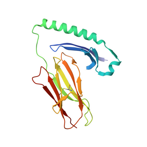
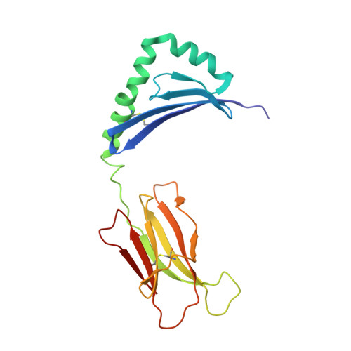
1R5V

Evidence that structural rearrangements and/or flexibility during TCR binding can contribute to T-cell activation


Experimental Data Snapshot

  • Method: X-RAY DIFFRACTION
  • Resolution: 2.50 Å
  • R-Value Free: 0.241 
  • R-Value Work: 0.213 
  • R-Value Observed: 0.215 

wwPDB Validation   3D Report Full Report


This is version 1.3 of the entry. See complete history


Literature

Evidence that structural rearrangements and/or flexibility during TCR binding can contribute to T cell activation.

Krogsgaard, M.Prado, N.Adams, E.J.He, X.L.Chow, D.C.Wilson, D.B.Garcia, K.C.Davis, M.M.

(2003) Mol Cell 12: 1367-1378

  • DOI: https://doi.org/10.1016/s1097-2765(03)00474-x
  • Primary Citation of Related Structures:  
    1R5V, 1R5W

  • PubMed Abstract: 

    While in many cases the half-life of T cell receptor (TCR) binding to a particular ligand is a good predictor of activation potential, numerous exceptions suggest that other physical parameter(s) must also play a role. Accordingly, we analyzed the thermodynamics of TCR binding to a series of peptide-MHC ligands, three of which are more stimulatory than their stability of binding would predict. Strikingly, we find that during TCR binding these outliers show anomalously large changes in heat capacity, an indicator of conformational change or flexibility in a binding interaction. By combining the values for heat capacity (DeltaCp) and the half-life of TCR binding (t(1/2)), we find that we can accurately predict the degree of T cell stimulation. Structural analysis shows significant changes in the central TCR contact residue of the peptide-MHC, indicating that structural rearrangements within the TCR-peptide-MHC interface can contribute to T cell activation.


  • Organizational Affiliation

    Howard Hughes Medical Institute, Stanford University School of Medicine, Stanford, CA 94305, USA.


Macromolecules
Find similar proteins by:  (by identity cutoff)  |  3D Structure
Entity ID: 1
MoleculeChains Sequence LengthOrganismDetailsImage
H-2 class II histocompatibility antigen, E-K alpha chainA,
D [auth C]
180Mus musculusMutation(s): 0 
UniProt
Find proteins for P04224 (Mus musculus)
Explore P04224 
Go to UniProtKB:  P04224
Entity Groups  
Sequence Clusters30% Identity50% Identity70% Identity90% Identity95% Identity100% Identity
UniProt GroupP04224
Sequence Annotations
Expand
  • Reference Sequence

Find similar proteins by:  Sequence   |   3D Structure  

Entity ID: 2
MoleculeChains Sequence LengthOrganismDetailsImage
artificial peptideB [auth E],
E [auth F]
13N/AMutation(s): 0 
Entity Groups  
Sequence Clusters30% Identity50% Identity70% Identity90% Identity95% Identity100% Identity
Sequence Annotations
Expand
  • Reference Sequence
Find similar proteins by:  (by identity cutoff)  |  3D Structure
Entity ID: 3
MoleculeChains Sequence LengthOrganismDetailsImage
MHC H2-IE-betaC [auth B],
F [auth D]
185Mus musculusMutation(s): 1 
UniProt & NIH Common Fund Data Resources
Find proteins for P18468 (Mus musculus)
Explore P18468 
Go to UniProtKB:  P18468
IMPC:  MGI:95901
Entity Groups  
Sequence Clusters30% Identity50% Identity70% Identity90% Identity95% Identity100% Identity
UniProt GroupP18468
Sequence Annotations
Expand
  • Reference Sequence
Experimental Data & Validation

Experimental Data

  • Method: X-RAY DIFFRACTION
  • Resolution: 2.50 Å
  • R-Value Free: 0.241 
  • R-Value Work: 0.213 
  • R-Value Observed: 0.215 
  • Space Group: P 21 21 21
Unit Cell:
Length ( Å )Angle ( ˚ )
a = 79.42α = 90
b = 104.65β = 90
c = 120.51γ = 90
Software Package:
Software NamePurpose
HKL-2000data collection
SCALEPACKdata scaling
MOLREPphasing
CNSrefinement
HKL-2000data reduction

Structure Validation

View Full Validation Report


Entry History 

Deposition Data

Revision History  (Full details and data files)

  • Version 1.0: 2004-02-03
    Type: Initial release
  • Version 1.1: 2008-04-29
    Changes: Version format compliance
  • Version 1.2: 2011-07-13
    Changes: Derived calculations, Version format compliance
  • Version 1.3: 2021-10-27
    Changes: Database references