Parallel Evolutionary Pathways for Glutathione Transferases: Structure and Mechanism of the Mitochondrial Class Kappa Enzyme rGSTK1-1
Ladner, J.E., Parsons, J.F., Rife, C.L., Gilliland, G.L., Armstrong, R.N.(2004) Biochemistry 43: 352-361
- PubMed: 14717589 Search on PubMed
- DOI: https://doi.org/10.1021/bi035832z
- Primary Citation Related Structures:
1R4W - PubMed Abstract:
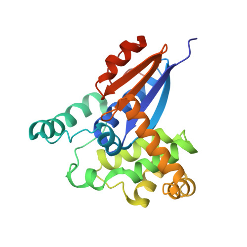
The class kappa glutathione (GSH) transferase is an enzyme that resides in the mitochondrial matrix. Its relationship to members of the canonical GSH transferase superfamily has remained an enigma. The three-dimensional structure of the class kappa enzyme from rat (rGSTK1-1) in complex with GSH has been solved by single isomorphous replacement with anomalous scattering at a resolution of 2.5 A. The structure reveals that the enzyme is more closely related to the protein disulfide bond isomerase, dsbA, from Escherichia coli than it is to members of the canonical superfamily. The structures of rGSTK1-1 and the canonical superfamily members indicate that the proteins folds have diverged from a common thioredoxin/glutaredoxin progenitor but did so by different mechanisms. The mitochondrial enzyme, therefore, represents a fourth protein superfamily that supports GSH transferase activity. The thioredoxin domain functions in a manner that is similar to that seen in the canonical enzymes by providing key structural elements for the recognition of GSH. The hydroxyl group of S16 is within hydrogen-bonding distance of the sulfur of bound GSH and is, in part, responsible for the ionization of the thiol in the E*GSH complex (pKa = 6.4 +/- 0.1). Preequilibrium kinetic experiments indicate that the k(on) for GSH is 1 x 10(5) M(-1) s(-1) and k(off) for GS- is approximately 8 s(-1) and relatively slow with respect to turnover with 1-chloro-2, 4-dinitrobenzene (CDNB). As a result, the KM(GSH) (11 mM) is much larger than the apparent Kd(GSH) (90 microM). The active site has a relatively open access channel that is flanked by disordered loops that may explain the relatively high turnover number (280 s(-1) at pH 7.0) toward CDNB. The disordered loops form an extensive contiguous patch on one face of the dimeric enzyme, a fact that suggests that the protein surface may interact with a membrane or other protein partner.
- The Center for Advanced Research in Biotechnology of the Maryland Biotechnology Institute and the National Institutes of Standards and Technology, Gudelsky Drive, Rockville, Maryland 20850, USA.
Organizational Affiliation: