


Crystal Structure of ADP-RIBOSYLTRANSFERASE C3bot2 from CLOSTRIDIUM BOTULINUM
Teplyakov, A., Obmolova, G., Gilliland, G.L., Narumiya, S.To be published.
Experimental Data Snapshot
Starting Model: experimental
View more details
wwPDB Validation 3D Report Full Report

Entity ID: 1 | |||||
|---|---|---|---|---|---|
| Molecule | Chains | Sequence Length | Organism | Details | Image |
| Mono-ADP-ribosyltransferase C3 | 204 | Clostridium phage c-st | Mutation(s): 0 EC: 2.4.2 | 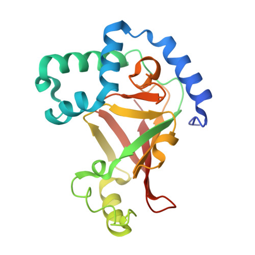 | |
UniProt | |||||
Entity Groups | |||||
| Sequence Clusters | 30% Identity50% Identity70% Identity90% Identity95% Identity100% Identity | ||||
| UniProt Group | Q00901 | ||||
Sequence AnnotationsExpand | |||||
Reference Sequence | |||||
| Length ( Å ) | Angle ( ˚ ) |
|---|---|
| a = 65.3 | α = 90 |
| b = 70 | β = 101.2 |
| c = 109 | γ = 90 |
| Software Name | Purpose |
|---|---|
| REFMAC | refinement |
| DENZO | data reduction |
| SCALEPACK | data scaling |
| AMoRE | phasing |


