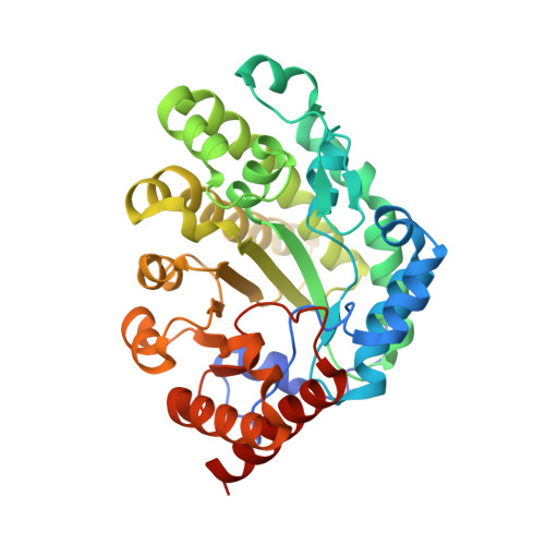
1R3Y

Uroporphyrinogen Decarboxylase in complex with coproporphyrinogen-III


Experimental Data Snapshot

  • Method: X-RAY DIFFRACTION
  • Resolution: 1.75 Å
  • R-Value Free: 0.186 
  • R-Value Work: 0.158 
  • R-Value Observed: 0.159 

Starting Model: experimental
View more details

wwPDB Validation   3D Report Full Report


Ligand Structure Quality Assessment 


This is version 1.4 of the entry. See complete history


Literature

Structural basis for tetrapyrrole coordination by uroporphyrinogen decarboxylase

Phillips, J.D.Whitby, F.G.Kushner, J.P.Hill, C.P.

(2003) EMBO J 22: 6225-6233

  • DOI: https://doi.org/10.1093/emboj/cdg606
  • Primary Citation of Related Structures:  
    1R3Q, 1R3R, 1R3S, 1R3T, 1R3V, 1R3W, 1R3Y

  • PubMed Abstract: 

    Uroporphyrinogen decarboxylase (URO-D), an essential enzyme that functions in the heme biosynthetic pathway, catalyzes decarboxylation of all four acetate groups of uroporphyrinogen to form coproporphyrinogen. Here we report crystal structures of URO-D in complex with the I and III isomer coproporphyrinogen products. Crystallization required use of a novel enzymatic approach to generate the highly oxygen-sensitive porphyrinogen substrate in situ. The tetrapyrrole product adopts a domed conformation that lies against a collar of conserved hydrophobic residues and allows formation of hydrogen bonding interactions between a carboxylate oxygen atom of the invariant Asp86 residue and the pyrrole NH groups. Structural and biochemical analyses of URO-D proteins mutated at Asp86 support the conclusion that this residue makes important contributions to binding and likely promotes catalysis by stabilizing a positive charge on a reaction intermediate. The central coordination geometry of Asp86 allows the initial substrates and the various partially decarboxylated intermediates to be bound with equivalent activating interactions, and thereby explains how all four of the substrate acetate groups can be decarboxylated at the same catalytic center.


  • Organizational Affiliation

    Department of Medicine, University of Utah School of Medicine, Salt Lake City, UT 84132, USA. john.phillips@hsc.utah.edu


Macromolecules
Find similar proteins by:  (by identity cutoff)  |  3D Structure
Entity ID: 1
MoleculeChains Sequence LengthOrganismDetailsImage
Uroporphyrinogen Decarboxylase367Homo sapiensMutation(s): 0 
EC: 4.1.1.37
UniProt & NIH Common Fund Data Resources
Find proteins for P06132 (Homo sapiens)
Explore P06132 
Go to UniProtKB:  P06132
PHAROS:  P06132
GTEx:  ENSG00000126088 
Entity Groups  
Sequence Clusters30% Identity50% Identity70% Identity90% Identity95% Identity100% Identity
UniProt GroupP06132
Sequence Annotations
Expand
  • Reference Sequence
Small Molecules
Ligands 1 Unique
IDChains Name / Formula / InChI Key2D Diagram3D Interactions
CP3
Query on CP3

Download Ideal Coordinates CCD File 
B [auth A]COPROPORPHYRINOGEN III
C36 H44 N4 O8
NIUVHXTXUXOFEB-UHFFFAOYSA-N
Experimental Data & Validation

Experimental Data

  • Method: X-RAY DIFFRACTION
  • Resolution: 1.75 Å
  • R-Value Free: 0.186 
  • R-Value Work: 0.158 
  • R-Value Observed: 0.159 
  • Space Group: P 31 2 1
Unit Cell:
Length ( Å )Angle ( ˚ )
a = 103.14α = 90
b = 103.14β = 90
c = 74.56γ = 120
Software Package:
Software NamePurpose
DENZOdata reduction
SCALEPACKdata scaling
REFMACrefinement

Structure Validation

View Full Validation Report


Ligand Structure Quality Assessment 


Entry History 

Deposition Data

Revision History  (Full details and data files)

  • Version 1.0: 2003-12-09
    Type: Initial release
  • Version 1.1: 2008-04-29
    Changes: Version format compliance
  • Version 1.2: 2011-07-13
    Changes: Version format compliance
  • Version 1.3: 2014-03-12
    Changes: Non-polymer description
  • Version 1.4: 2023-08-23
    Changes: Data collection, Database references, Derived calculations, Refinement description