1QZ5

Structure of rabbit actin in complex with kabiramide C


Experimental Data Snapshot

  • Method: X-RAY DIFFRACTION
  • Resolution: 1.45 Å
  • R-Value Free: 0.187 
  • R-Value Work: 0.169 
  • R-Value Observed: 0.170 

wwPDB Validation   3D Report Full Report


Ligand Structure Quality Assessment 


This is version 1.2 of the entry. See complete history


Literature

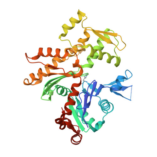
Trisoxazole macrolide toxins mimic the binding of actin-capping proteins to actin

Klenchin, V.A.Allingham, J.S.King, R.Tanaka, J.Marriott, G.Rayment, I.

(2003) Nat Struct Biol 10: 1058-1063

  • DOI: https://doi.org/10.1038/nsb1006
  • Primary Citation of Related Structures:  
    1QZ5, 1QZ6

  • PubMed Abstract: 
  • Marine macrolide toxins of trisoxazole family target actin with high affinity and specificity and have promising pharmacological properties. We present X-ray structures of actin in complex with two members of this family, kabiramide C and jaspisamide A, at a resolution of 1 ...

    Marine macrolide toxins of trisoxazole family target actin with high affinity and specificity and have promising pharmacological properties. We present X-ray structures of actin in complex with two members of this family, kabiramide C and jaspisamide A, at a resolution of 1.45 and 1.6 A, respectively. The structures reveal the absolute stereochemistry of these toxins and demonstrate that their trisoxazole ring interacts with actin subdomain 1 while the aliphatic side chain is inserted into the hydrophobic cavity between actin subdomains 1 and 3. The binding site is essentially the same as the one occupied by the actin-capping domain of the gelsolin superfamily of proteins. The structural evidence suggests that actin filament severing and capping by these toxins is also analogous to that of gelsolin. Consequently, these macrolides may be viewed as small molecule biomimetics of an entire class of actin-binding proteins.


    Organizational Affiliation

    Department of Biochemistry, University of Wisconsin at Madison, 433 Babcock Drive, Madison, Wisconsin 53706, USA.


Macromolecules
Find similar proteins by:  (by identity cutoff)  |  3D Structure
Entity ID: 1
MoleculeChainsSequence LengthOrganismDetailsImage
Actin, alpha skeletal muscle375Oryctolagus cuniculusMutation(s): 1 
Gene Names: ACTA1ACTA
UniProt
Find proteins for P68135 (Oryctolagus cuniculus)
Explore P68135 
Go to UniProtKB:  P68135
Entity Groups  
Sequence Clusters30% Identity50% Identity70% Identity90% Identity95% Identity100% Identity
UniProt GroupP68135
Protein Feature View
Expand
  • Reference Sequence
Small Molecules
Ligands 3 Unique
IDChainsName / Formula / InChI Key2D Diagram3D Interactions
KAB
Query on KAB

Download Ideal Coordinates CCD File 
D [auth A]KABIRAMIDE C
C48 H75 N5 O14
XYKNXOJYKRVXBX-ZAUPHERQSA-N
 Ligand Interaction
ATP
Query on ATP

Download Ideal Coordinates CCD File 
C [auth A]ADENOSINE-5'-TRIPHOSPHATE
C10 H16 N5 O13 P3
ZKHQWZAMYRWXGA-KQYNXXCUSA-N
 Ligand Interaction
CA
Query on CA

Download Ideal Coordinates CCD File 
B [auth A]CALCIUM ION
Ca
BHPQYMZQTOCNFJ-UHFFFAOYSA-N
 Ligand Interaction
Modified Residues  1 Unique
IDChainsTypeFormula2D DiagramParent
HIC
Query on HIC
A
L-PEPTIDE LINKINGC7 H11 N3 O2HIS
Experimental Data & Validation

Experimental Data

  • Method: X-RAY DIFFRACTION
  • Resolution: 1.45 Å
  • R-Value Free: 0.187 
  • R-Value Work: 0.169 
  • R-Value Observed: 0.170 
  • Space Group: P 21 21 21
Unit Cell:
Length ( Å )Angle ( ˚ )
a = 69.919α = 90
b = 70.617β = 90
c = 75.13γ = 90
Software Package:
Software NamePurpose
REFMACrefinement
HKL-2000data reduction
SCALEPACKdata scaling
MOLREPphasing

Structure Validation

View Full Validation Report


Ligand Structure Quality Assessment 


Entry History 

Deposition Data

Revision History  (Full details and data files)

  • Version 1.0: 2003-11-11
    Type: Initial release
  • Version 1.1: 2008-04-29
    Changes: Version format compliance
  • Version 1.2: 2011-07-13
    Changes: Version format compliance