1QX1

Golgi alpha-mannosidase II D341N mutant complex with 2-F-mannosyl-F


Experimental Data Snapshot

  • Method: X-RAY DIFFRACTION
  • Resolution: 1.30 Å
  • R-Value Free: 0.189 
  • R-Value Work: 0.170 
  • R-Value Observed: 0.170 

wwPDB Validation   3D Report Full Report


Ligand Structure Quality Assessment 


This is version 2.2 of the entry. See complete history


Literature

Insights into the mechanism of Drosophila melanogaster Golgi alpha-mannosidase II through the structural analysis of covalent reaction intermediates.

Numao, S.Kuntz, D.A.Withers, S.G.Rose, D.R.

(2003) J Biol Chem 278: 48074-48083

  • DOI: https://doi.org/10.1074/jbc.M309249200
  • Primary Citation of Related Structures:  
    1QWN, 1QWU, 1QX1

  • PubMed Abstract: 

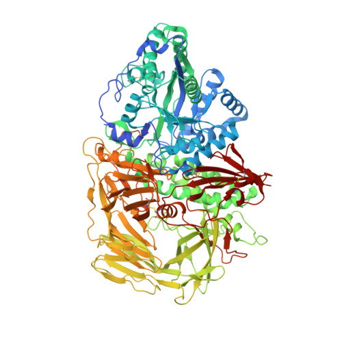
    The family 38 golgi alpha-mannosidase II, thought to cleave mannosidic bonds through a double displacement mechanism involving a reaction intermediate, is a clinically important enzyme involved in glycoprotein processing. The structure of three different covalent glycosyl-enzyme intermediates have been determined to 1.2-A resolution for the Golgi alpha-mannosidase II from Drosophila melanogaster by use of fluorinated sugar analogues, both with the wild-type enzyme and a mutant enzyme in which the acid/base catalyst has been removed. All these structures reveal sugar intermediates bound in a distorted 1S5 skew boat conformation. The similarity of this conformation with that of the substrate in the recently determined structure of the Michaelis complex of a beta-mannanase (Ducros, V. M. A., Zechel, D. L., Murshudov, G. N., Gilbert, H. J., Szabo, L., Stoll, D., Withers, S. G., and Davies, G. J. (2002) Angew. Chem. Int. Ed. Engl. 41, 2824-2827) suggests that these disparate enzymes have recruited common stereoelectronic features in evolving their catalytic mechanisms.


  • Organizational Affiliation

    Department of Chemistry, University of British Columbia, Vancouver, British Columbia V6T 1Z1, Canada.


Macromolecules
Find similar proteins by:  (by identity cutoff)  |  3D Structure
Entity ID: 1
MoleculeChains Sequence LengthOrganismDetailsImage
Alpha-mannosidase II1,045Drosophila melanogasterMutation(s): 1 
Gene Names: ALPHA-MAN-II OR GMII OR CG18474/CG18802/CG8139
EC: 3.2.1.114
UniProt
Find proteins for Q24451 (Drosophila melanogaster)
Explore Q24451 
Go to UniProtKB:  Q24451
Entity Groups  
Sequence Clusters30% Identity50% Identity70% Identity90% Identity95% Identity100% Identity
UniProt GroupQ24451
Sequence Annotations
Expand
  • Reference Sequence
Small Molecules
Binding Affinity Annotations 
IDSourceBinding Affinity
GUL PDBBind:  1QX1 Ki: 7.50e+6 (nM) from 1 assay(s)
Experimental Data & Validation

Experimental Data

  • Method: X-RAY DIFFRACTION
  • Resolution: 1.30 Å
  • R-Value Free: 0.189 
  • R-Value Work: 0.170 
  • R-Value Observed: 0.170 
  • Space Group: P 21 21 21
Unit Cell:
Length ( Å )Angle ( ˚ )
a = 69.046α = 90
b = 109.826β = 90
c = 138.907γ = 90
Software Package:
Software NamePurpose
CNSrefinement
DENZOdata reduction
SCALEPACKdata scaling
CNSphasing

Structure Validation

View Full Validation Report


Ligand Structure Quality Assessment 


Entry History 

Deposition Data

Revision History  (Full details and data files)

  • Version 1.0: 2003-10-07
    Type: Initial release
  • Version 1.1: 2008-04-29
    Changes: Version format compliance
  • Version 1.2: 2011-07-13
    Changes: Non-polymer description, Version format compliance
  • Version 2.0: 2020-07-29
    Type: Remediation
    Reason: Carbohydrate remediation
    Changes: Advisory, Atomic model, Data collection, Database references, Derived calculations, Structure summary
  • Version 2.1: 2021-10-27
    Changes: Database references, Structure summary
  • Version 2.2: 2023-08-23
    Changes: Data collection, Refinement description