Conservation of structure and function among histidine-containing phosphotransfer (HPt) domains as revealed by the crystal structure of YPD1.
Xu, Q., West, A.H.(1999) J Mol Biol 292: 1039-1050
- PubMed: 10512701 Search on PubMed
- DOI: https://doi.org/10.1006/jmbi.1999.3143
- Primary Citation Related Structures:
1QSP - PubMed Abstract:
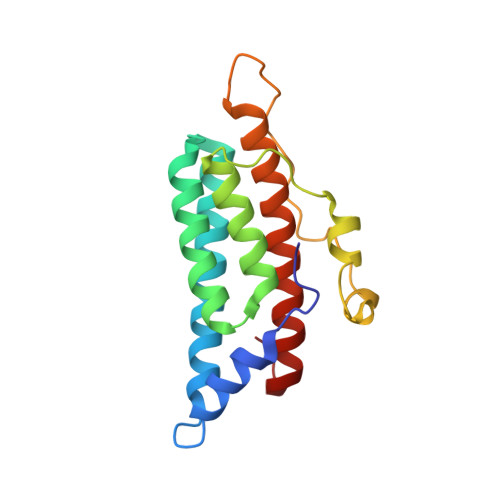
In Saccharomyces cerevisiae, the SLN1-YPD1-SSK1 phosphorelay system controls a downstream mitogen-activated protein (MAP) kinase in response to hyperosmotic stress. YPD1 functions as a phospho-histidine protein intermediate which is required for phosphoryl group transfer from the sensor kinase SLN1 to the response regulator SSK1. In addition, YPD1 mediates phosphoryl transfer from SLN1 to SKN7, the only other response regulator protein in yeast which plays a role in response to oxidative stress and cell wall biosynthesis. The X-ray structure of YPD1 was solved at a resolution of 2.7 A by conventional multiple isomorphous replacement with anomalous scattering. The tertiary structure of YPD1 consists of six alpha-helices and a short 310-helix. A four-helix bundle comprises the central core of the molecule and contains the histidine residue that is phosphorylated. Structure-based comparisons of YPD1 to other proteins having a similar function, such as the Escherichia coli ArcB histidine-containing phosphotransfer (HPt) domain and the P1 domain of the CheA kinase, revealed that the helical bundle and several structural features around the active-site histidine residue are conserved between the prokaryotic and eukaryotic kingdoms. Despite limited amino acid sequence homology among HPt domains, our analysis of YPD1 as a prototypical family member, indicates that these phosphotransfer domains are likely to share a similar fold and common features with regard to response regulator binding and mechanism for histidine-aspartate phosphoryl transfer.
- Department of Chemistry and Biochemistry, University of Oklahoma, 620 Parrington Oval, Norman, OK, USA.
Organizational Affiliation: