1QS4 | pdb_00001qs4

Core domain of HIV-1 integrase complexed with Mg++ and 1-(5-chloroindol-3-yl)-3-hydroxy-3-(2H-tetrazol-5-yl)-propenone


Experimental Data Snapshot

  • Method: X-RAY DIFFRACTION
  • Resolution: 2.10 Å
  • R-Value Free: 
    0.255 (Depositor) 
  • R-Value Work: 
    0.209 (Depositor) 
  • R-Value Observed: 
    0.209 (Depositor) 

wwPDB Validation 3D Report Full Report

Validation slider image for 1QS4

This is version 1.5 of the entry. See complete history

Literature

Structure of the HIV-1 integrase catalytic domain complexed with an inhibitor: a platform for antiviral drug design.

Goldgur, Y.Craigie, R.Cohen, G.H.Fujiwara, T.Yoshinaga, T.Fujishita, T.Sugimoto, H.Endo, T.Murai, H.Davies, D.R.

(1999) Proc Natl Acad Sci U S A 96: 13040-13043

  • DOI: https://doi.org/10.1073/pnas.96.23.13040
  • Primary Citation Related Structures: 
    1QS4

  • PubMed Abstract: 

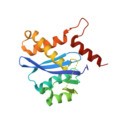
    HIV integrase, the enzyme that inserts the viral DNA into the host chromosome, has no mammalian counterpart, making it an attractive target for antiviral drug design. As one of the three enzymes produced by HIV, it can be expected that inhibitors of this enzyme will complement the therapeutic use of HIV protease and reverse transcriptase inhibitors. We have determined the structure of a complex of the HIV-1 integrase core domain with a novel inhibitor, 5ClTEP, 1-(5-chloroindol-3-yl)-3-hydroxy-3-(2H-tetrazol-5-yl)-pro penone, to 2.1-A resolution. The inhibitor binds centrally in the active site of the integrase and makes a number of close contacts with the protein. Only minor changes in the protein accompany inhibitor binding. This inhibitor complex will provide a platform for structure-based design of an additional class of inhibitors for antiviral therapy.


  • Organizational Affiliation
    • Laboratory of Molecular Biology, National Institute of Diabetes, Digestive and Kidney Diseases, National Institutes of Health, Bethesda, MD 20892-0560, USA.

Macromolecule Content 

  • Total Structure Weight: 50.8 kDa 
  • Atom Count: 4,036 
  • Modeled Residue Count: 453 
  • Deposited Residue Count: 462 
  • Unique protein chains: 1

Macromolecules

Find similar proteins by:|  3D Structure
Entity ID: 1
MoleculeChains  Sequence LengthOrganismDetailsImage
PROTEIN (HIV-1 INTEGRASE (E.C.2.7.7.49))
A, B, C
154Human immunodeficiency virus 1Mutation(s): 2 
EC: 2.7.7.49
UniProt
Find proteins for P12497 (Human immunodeficiency virus type 1 group M subtype B (isolate NY5))
Explore P12497 
Go to UniProtKB:  P12497
Entity Groups
Sequence Clusters30% Identity50% Identity70% Identity90% Identity95% Identity100% Identity
UniProt GroupP12497
Sequence Annotations
Expand
Reference Sequence

Experimental Data & Validation

Experimental Data

  • Method: X-RAY DIFFRACTION
  • Resolution: 2.10 Å
  • R-Value Free:  0.255 (Depositor) 
  • R-Value Work:  0.209 (Depositor) 
  • R-Value Observed: 0.209 (Depositor) 
Space Group: C 1 2 1
Unit Cell:
Length ( Å )Angle ( ˚ )
a = 76.233α = 90
b = 46.735β = 105.55
c = 141.361γ = 90
Software Package:
Software NamePurpose
AMoREphasing
CNSrefinement
DENZOdata reduction
SCALEPACKdata scaling

Structure Validation

View Full Validation Report


Entry History 

Deposition Data

Revision History  (Full details and data files)

  • Version 1.0: 1999-11-17
    Type: Initial release
  • Version 1.1: 2008-03-26
    Changes: Version format compliance
  • Version 1.2: 2011-07-13
    Changes: Derived calculations, Version format compliance
  • Version 1.3: 2017-02-01
    Changes: Structure summary
  • Version 1.4: 2021-11-03
    Changes: Database references, Derived calculations
  • Version 1.5: 2024-02-14
    Changes: Data collection