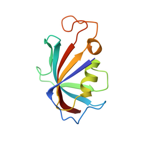
1QPF

FK506 BINDING PROTEIN (12 KDA, HUMAN) COMPLEX WITH L-709,858


Experimental Data Snapshot

  • Method: X-RAY DIFFRACTION
  • Resolution: 2.50 Å
  • R-Value Free: 0.310 
  • R-Value Work: 0.243 
  • R-Value Observed: 0.243 

Starting Model: experimental
View more details

wwPDB Validation   3D Report Full Report


Ligand Structure Quality Assessment 


This is version 1.5 of the entry. See complete history


Literature

32-Indolyl ether derivatives of ascomycin: three-dimensional structures of complexes with FK506-binding protein.

Becker, J.W.Rotonda, J.Cryan, J.G.Martin, M.Parsons, W.H.Sinclair, P.J.Wiederrecht, G.Wong, F.

(1999) J Med Chem 42: 2798-2804

  • DOI: https://doi.org/10.1021/jm9806042
  • Primary Citation of Related Structures:  
    1QPF, 1QPL

  • PubMed Abstract: 

    32-Indole ether derivatives of tacrolimus and ascomycin retain the potent immunosuppressive activity of their parent compounds but display reduced toxicity. In addition, their complexes with the 12-kDa FK506-binding protein (FKBP) form more stable complexes with the protein phosphatase calcineurin, the molecular target of these drugs. We have solved the three-dimensional structures of the FKBP complexes with two 32-indolyl derivatives of ascomycin. The structures of the protein and the macrolide are remarkably similar to those seen in the complexes with tacrolimus and ascomycin. The indole groups project away from the body of the complex, and multiple conformations are observed for the linkage to these groups as well as for a nearby peptide suggesting apparent flexibility in these parts of the structure. Comparison of these structures with that of the ternary complex of calcineurin, FKBP, and tacrolimus suggests that the indole groups interact with a binding site comprising elements of both the calcineurin alpha- and beta-chains and that this interaction is responsible for the increased stability of these complexes.


  • Organizational Affiliation

    Departments of Endocrinology and Chemical Biology, Medicinal Chemistry, and Immunology Research, Merck Research Laboratories, P.O. Box 2000, Rahway, New Jersey 07065-0900, USA. beckerj@merck.com


Macromolecules
Find similar proteins by:  (by identity cutoff)  |  3D Structure
Entity ID: 1
MoleculeChains Sequence LengthOrganismDetailsImage
PROTEIN (FK506-BINDING PROTEIN)A,
B [auth D]
107Homo sapiensMutation(s): 0 
UniProt & NIH Common Fund Data Resources
Find proteins for P62942 (Homo sapiens)
Explore P62942 
Go to UniProtKB:  P62942
PHAROS:  P62942
GTEx:  ENSG00000088832 
Entity Groups  
Sequence Clusters30% Identity50% Identity70% Identity90% Identity95% Identity100% Identity
UniProt GroupP62942
Sequence Annotations
Expand
  • Reference Sequence
Small Molecules
Binding Affinity Annotations 
IDSourceBinding Affinity
858 PDBBind:  1QPF IC50: 27 (nM) from 1 assay(s)
Experimental Data & Validation

Experimental Data

  • Method: X-RAY DIFFRACTION
  • Resolution: 2.50 Å
  • R-Value Free: 0.310 
  • R-Value Work: 0.243 
  • R-Value Observed: 0.243 
  • Space Group: P 61 2 2
Unit Cell:
Length ( Å )Angle ( ˚ )
a = 74.26α = 90
b = 74.26β = 90
c = 236.45γ = 120
Software Package:
Software NamePurpose
X-PLORmodel building
X-PLORrefinement
X-GENdata scaling
X-PLORphasing

Structure Validation

View Full Validation Report


Ligand Structure Quality Assessment 


Entry History 

Deposition Data

Revision History  (Full details and data files)

  • Version 1.0: 1999-08-16
    Type: Initial release
  • Version 1.1: 2008-04-27
    Changes: Version format compliance
  • Version 1.2: 2011-07-13
    Changes: Derived calculations, Version format compliance
  • Version 1.3: 2017-10-04
    Changes: Refinement description
  • Version 1.4: 2020-07-29
    Type: Remediation
    Reason: Carbohydrate remediation
    Changes: Data collection, Derived calculations, Structure summary
  • Version 1.5: 2023-08-16
    Changes: Data collection, Database references, Refinement description, Structure summary