Crystal Structure of Avian Carboxypeptidase D Domain II : A Prototype for the Regulatory Metallocarboxypeptidase Subfamily
Gomis-Rueth, F.X., Companys, V., Qian, Y., Fricker, L.D., Vendrell, J., Aviles, F.X., Coll, M.(1999) EMBO J 18: 5817
- PubMed: 10545093 Search on PubMedSearch on PubMed Central
- DOI: https://doi.org/10.1093/emboj/18.21.5817
- Primary Citation Related Structures:
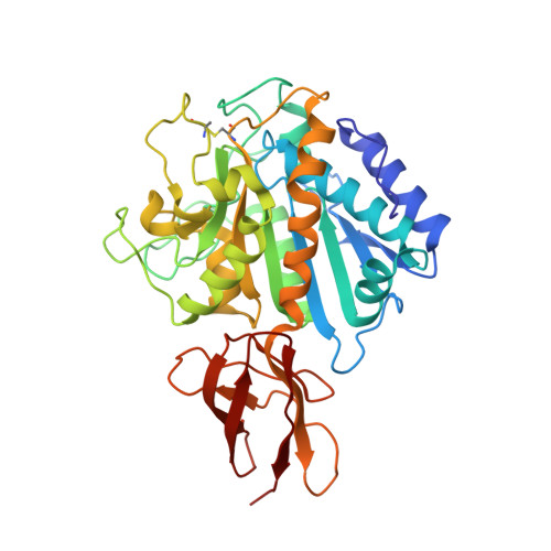
1QMU - PubMed Abstract:
The crystal structure of domain II of duck carboxypeptidase D, a prohormone/propeptide processing enzyme integrated in a three repeat tandem in the natural system, has been solved, constituting a prototype for members of the regulatory metallocarboxypeptidase subfamily. It displays a 300 residue N-terminal alpha/beta-hydrolase subdomain with overall topological similarity to and general coincidence of the key catalytic residues with the archetypal pancreatic carboxypeptidase A. However, numerous significant insertions/deletions in segments forming the funnel-like access to the active site explain differences in specificity towards larger protein substrates or inhibitors. This alpha/beta-hydrolase subdomain is followed by a C-terminal 80 residue beta-sandwich subdomain, unique for these regulatory metalloenzymes and topologically related to transthyretin and sugar-binding proteins. The structure described here establishes the fundamentals for a better understanding of the mechanism ruling events such as prohormone processing and will enable modelling of regulatory carboxypeptidases as well as a more rational design of inhibitors of carboxypeptidase D.
- Institut de Biologia Molecular de Barcelona, CID-CSIC, Jordi Girona, 18-26, 08034 Barcelona, Spain.
Organizational Affiliation: