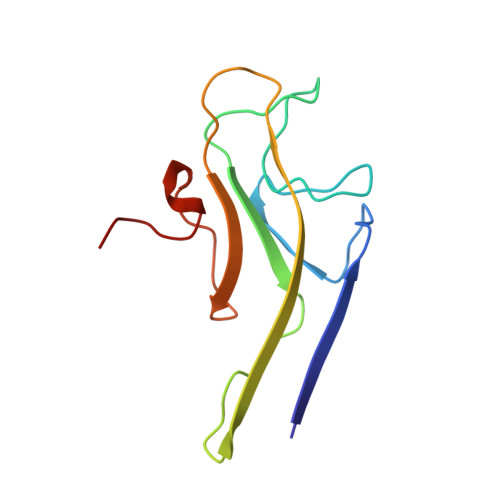
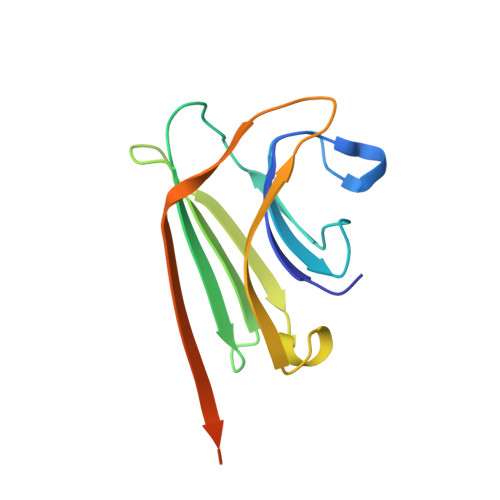
1QMO | pdb_00001qmo

Structure of FRIL, a legume lectin that delays hematopoietic progenitor maturation


Experimental Data Snapshot

  • Method: X-RAY DIFFRACTION
  • Resolution: 3.50 Å
  • R-Value Free: 
    0.252 (Depositor), 0.250 (DCC) 
  • R-Value Work: 
    0.232 (Depositor), 0.230 (DCC) 
  • R-Value Observed: 
    0.232 (Depositor) 

wwPDB Validation   3D Report Full Report


Ligand Structure Quality Assessment 

Created with Raphaël 2.3.0Worse 01 BetterLigand structure goodness of fit to experimental dataBest fitted MANClick on this verticalbar to view details

This is version 1.4 of the entry. See complete history


Literature

The Role of Weak Protein-Protein Interactions in Multivalent Lectin-Carbohydrate Binding: Crystal Structure of Cross-Linked Fril

Hamelryck, T.W.Moore, J.G.Chrispeels, M.J.Loris, R.Wyns, L.

(2000) J Mol Biology 299: 875

  • DOI: https://doi.org/10.1006/jmbi.2000.3785
  • Primary Citation of Related Structures:  
    1QMO

  • PubMed Abstract: 

    Binding of multivalent glycoconjugates by lectins often leads to the formation of cross-linked complexes. Type I cross-links, which are one-dimensional, are formed by a divalent lectin and a divalent glycoconjugate. Type II cross-links, which are two or three-dimensional, occur when a lectin or glycoconjugate has a valence greater than two. Type II complexes are a source of additional specificity, since homogeneous type II complexes are formed in the presence of mixtures of lectins and glycoconjugates. This additional specificity is thought to become important when a lectin interacts with clusters of glycoconjugates, e.g. as is present on the cell surface. The cryst1al structure of the Glc/Man binding legume lectin FRIL in complex with a trisaccharide provides a molecular snapshot of how weak protein-protein interactions, which are not observed in solution, can become important when a cross-linked complex is formed. In solution, FRIL is a divalent dimer, but in the crystal FRIL forms a tetramer, which allows for the formation of an intricate type II cross-linked complex with the divalent trisaccharide. The dependence on weak protein-protein interactions can ensure that a specific type II cross-linked complex and its associated specificity can occur only under stringent conditions, which explains why lectins are often found forming higher-order oligomers.


  • Organizational Affiliation

    Laboratorium voor Ultrastructuur, Vlaams Interuniversitair Instituut voor Biotechnologie, Vrije Universiteit Brussel, Paardenstraat 65, Sint-Genesius-Rode, B-1640, Belgium. thamelry@vub.ac.be


Macromolecules
Find similar proteins by:  (by identity cutoff)  |  3D Structure
Entity ID: 1
MoleculeChains Sequence LengthOrganismDetailsImage
MANNOSE BINDING LECTIN, FRIL
A, B, C, D
113Lablab purpureusMutation(s): 0 
UniProt
Find proteins for Q9ZTA9 (Lablab purpureus)
Explore Q9ZTA9 
Go to UniProtKB:  Q9ZTA9
Entity Groups  
Sequence Clusters30% Identity50% Identity70% Identity90% Identity95% Identity100% Identity
UniProt GroupQ9ZTA9
Sequence Annotations
Expand
  • Reference Sequence
Find similar proteins by:  (by identity cutoff)  |  3D Structure
Entity ID: 2
MoleculeChains Sequence LengthOrganismDetailsImage
MANNOSE BINDING LECTIN, FRIL
E, F, G, H
133Lablab purpureusMutation(s): 0 
UniProt
Find proteins for Q9ZTA9 (Lablab purpureus)
Explore Q9ZTA9 
Go to UniProtKB:  Q9ZTA9
Entity Groups  
Sequence Clusters30% Identity50% Identity70% Identity90% Identity95% Identity100% Identity
UniProt GroupQ9ZTA9
Sequence Annotations
Expand
  • Reference Sequence
Small Molecules
Ligands 3 Unique
IDChains Name / Formula / InChI Key2D Diagram3D Interactions
MAN
Query on MAN

Download Ideal Coordinates CCD File 
K [auth A],
N [auth B],
Q [auth C],
T [auth D]
alpha-D-mannopyranose
C6 H12 O6
WQZGKKKJIJFFOK-PQMKYFCFSA-N
MN
Query on MN

Download Ideal Coordinates CCD File 
J [auth A],
M [auth B],
P [auth C],
S [auth D]
MANGANESE (II) ION
Mn
WAEMQWOKJMHJLA-UHFFFAOYSA-N
CA
Query on CA

Download Ideal Coordinates CCD File 
I [auth A],
L [auth B],
O [auth C],
R [auth D]
CALCIUM ION
Ca
BHPQYMZQTOCNFJ-UHFFFAOYSA-N
Experimental Data & Validation

Experimental Data

  • Method: X-RAY DIFFRACTION
  • Resolution: 3.50 Å
  • R-Value Free:  0.252 (Depositor), 0.250 (DCC) 
  • R-Value Work:  0.232 (Depositor), 0.230 (DCC) 
  • R-Value Observed: 0.232 (Depositor) 
Space Group: P 65 2 2
Unit Cell:
Length ( Å )Angle ( ˚ )
a = 151.36α = 90
b = 151.36β = 90
c = 309.88γ = 120
Software Package:
Software NamePurpose
CNSrefinement
DENZOdata reduction
SCALEPACKdata scaling
CNSphasing

Structure Validation

View Full Validation Report


Ligand Structure Quality Assessment 

Created with Raphaël 2.3.0Worse 01 BetterLigand structure goodness of fit to experimental dataBest fitted MANClick on this verticalbar to view details

Entry History 

Deposition Data

Revision History  (Full details and data files)

  • Version 1.0: 1999-10-10
    Type: Initial release
  • Version 1.1: 2014-09-03
    Changes: Database references, Derived calculations, Version format compliance
  • Version 1.2: 2019-07-24
    Changes: Data collection
  • Version 1.3: 2020-07-29
    Type: Remediation
    Reason: Carbohydrate remediation
    Changes: Data collection, Derived calculations, Other, Structure summary
  • Version 1.4: 2024-05-08
    Changes: Data collection, Database references, Structure summary