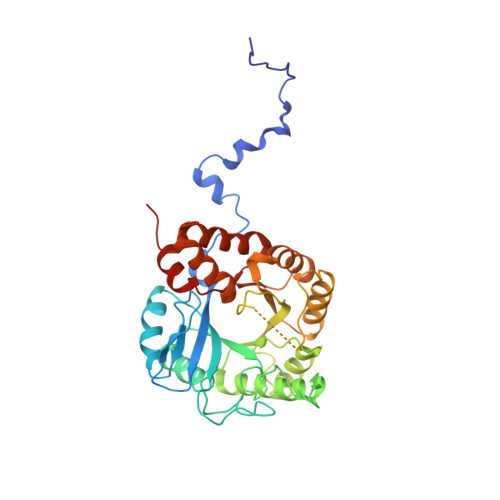
1QML

Hg complex of yeast 5-aminolaevulinic acid dehydratase


Experimental Data Snapshot

  • Method: X-RAY DIFFRACTION
  • Resolution: 3.00 Å
  • R-Value Free: 0.330 
  • R-Value Work: 0.249 

Starting Model: experimental
View more details

wwPDB Validation   3D Report Full Report


This is version 1.4 of the entry. See complete history


Literature

MAD Analyses of Yeast 5-Aminolaevulinic Acid Dehydratase. Their Use in Structure Determination and in Defining the Metal Binding Sites

Erskine, P.T.Duke, E.M.H.Tickle, I.J.Senior, N.M.Warren, M.J.Cooper, J.B.

(2000) Acta Crystallogr D Biol Crystallogr 56: 421

  • DOI: https://doi.org/10.1107/s0907444900000597
  • Primary Citation of Related Structures:  
    1QML, 1QNV

  • PubMed Abstract: 

    MAD experiments attempting to solve the structure of 5--aminolaevulinic acid dehydratase using Zn and Pb edges are described. The data obtained proved insufficient for a complete structure solution but were invaluable in subsequent identification of metal-binding sites using anomalous difference Fourier analyses once the structure of the enzyme had been solved. These sites include the highly inhibitory substitution of an enzymic cofactor Zn(2+) ion by Pb(2+) ions, which represents a major contribution towards understanding the molecular basis of lead poisoning. The MAD data collected at the Pb edge were also used with isomorphous replacement data from the same Pb co-crystal and a Hg co-crystal to provide the first delineation of the enzyme's quaternary structure. In this MADIR analysis, the Hg co-crystal data were treated as native data. Anomalous difference Fouriers were again used, revealing that Hg(2+) had substituted for the same Zn(2+) cofactor ion as had Pb(2+), a finding of fundamental importance for the understanding of mercury poisoning. In addition, Pt(2+) ions were found to bind at the same place in the structure. The refined structures of the Pb- and the Hg-complexed enzymes are presented at 2.5 and 3.0 A resolution, respectively.


  • Organizational Affiliation

    Biochemistry and Molecular Biology, School of Biological Sciences, University of Southampton, Bassett Crescent East, Southampton SO16 7PX, England.


Macromolecules
Find similar proteins by:  (by identity cutoff)  |  3D Structure
Entity ID: 1
MoleculeChains Sequence LengthOrganismDetailsImage
5-AMINOLAEVULINIC ACID DEHYDRATASE342Saccharomyces cerevisiaeMutation(s): 0 
EC: 4.2.1.24
UniProt
Find proteins for P05373 (Saccharomyces cerevisiae (strain ATCC 204508 / S288c))
Explore P05373 
Go to UniProtKB:  P05373
Entity Groups  
Sequence Clusters30% Identity50% Identity70% Identity90% Identity95% Identity100% Identity
UniProt GroupP05373
Sequence Annotations
Expand
  • Reference Sequence
Small Molecules
Ligands 1 Unique
IDChains Name / Formula / InChI Key2D Diagram3D Interactions
HG
Query on HG

Download Ideal Coordinates CCD File 
B [auth A]MERCURY (II) ION
Hg
BQPIGGFYSBELGY-UHFFFAOYSA-N
Experimental Data & Validation

Experimental Data

  • Method: X-RAY DIFFRACTION
  • Resolution: 3.00 Å
  • R-Value Free: 0.330 
  • R-Value Work: 0.249 
  • Space Group: I 4 2 2
Unit Cell:
Length ( Å )Angle ( ˚ )
a = 102.05α = 90
b = 102.05β = 90
c = 170.49γ = 90
Software Package:
Software NamePurpose
CCP4refinement
MOSFLMdata reduction
Agrovatadata scaling
SCALAdata scaling
CCP4phasing

Structure Validation

View Full Validation Report


Entry History 

Deposition Data

Revision History  (Full details and data files)

  • Version 1.0: 2000-10-06
    Type: Initial release
  • Version 1.1: 2011-05-08
    Changes: Version format compliance
  • Version 1.2: 2011-07-13
    Changes: Version format compliance
  • Version 1.3: 2019-05-22
    Changes: Data collection, Other, Refinement description
  • Version 1.4: 2023-12-13
    Changes: Data collection, Database references, Derived calculations, Other, Refinement description