The 2.2 A structure of the rRNA methyltransferase ErmC' and its complexes with cofactor and cofactor analogs: implications for the reaction mechanism.
Schluckebier, G., Zhong, P., Stewart, K.D., Kavanaugh, T.J., Abad-Zapatero, C.(1999) J Mol Biology 289: 277-291
- PubMed: 10366505 Search on PubMed
- DOI: https://doi.org/10.1006/jmbi.1999.2788
- Primary Citation Related Structures:
1QAM, 1QAN, 1QAO, 1QAQ - PubMed Abstract:
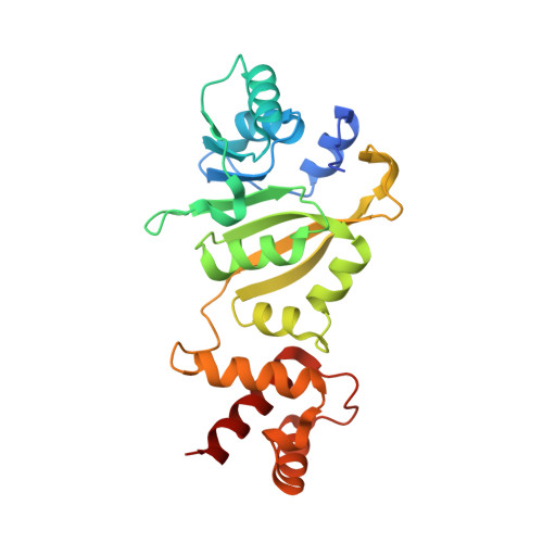
The rRNA methyltransferase ErmC' transfers methyl groups from S -adenosyl-l-methionine to atom N6 of an adenine base within the peptidyltransferase loop of 23 S rRNA, thus conferring antibiotic resistance against a number of macrolide antibiotics. The crystal structures of ErmC' and of its complexes with the cofactor S -adenosyl-l-methionine, the reaction product S-adenosyl-l-homocysteine and the methyltransferase inhibitor Sinefungin, respectively, show that the enzyme undergoes small conformational changes upon ligand binding. Overall, the ligand molecules bind to the protein in a similar mode as observed for other methyltransferases. Small differences between the binding of the amino acid parts of the different ligands are correlated with differences in their chemical structure. A model for the transition-state based on the atomic details of the active site is consistent with a one-step methyl-transfer mechanism and might serve as a first step towards the design of potent Erm inhibitors.
- Abbott Laboratories, D46Y-AP 10, 100 Abbott Park Road, Abbott Park, IL, 60064, USA. gerd.schluckebier@abbott.com
Organizational Affiliation: