1QA4

HIV-1 NEF ANCHOR DOMAIN, NMR, 2 STRUCTURES


Experimental Data Snapshot

  • Method: SOLUTION NMR
  • Conformers Calculated: 400 
  • Conformers Submitted: 
  • Selection Criteria: TWO STRUCTURES WITH LOW TOTAL ENERGY WERE SELECTED SHOWING THE CONFORMATIONAL VARIETY OF THE FLEXIBLE DOMAIN 

wwPDB Validation   3D Report Full Report

Currently 1QA4 does not have a validation slider image.


This is version 1.4 of the entry. See complete history


Literature

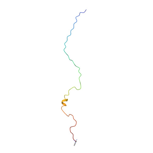
Structure of the anchor-domain of myristoylated and non-myristoylated HIV-1 Nef protein.

Geyer, M.Munte, C.E.Schorr, J.Kellner, R.Kalbitzer, H.R.

(1999) J Mol Biol 289: 123-138

  • DOI: https://doi.org/10.1006/jmbi.1999.2740
  • Primary Citation of Related Structures:  
    1QA4, 1QA5

  • PubMed Abstract: 
  • Negative factor (Nef) is a regulatory myristoylated protein of human immunodeficiency virus (HIV) that has a two-domain structure consisting of an anchor domain and a core domain separated by a specific cleavage site of the HIV proteases. For structural analysis, the HIV-1 Nef anchor domain (residues 2-57) was synthesized with a myristoylated and non-myristoylated N terminus ...

    Negative factor (Nef) is a regulatory myristoylated protein of human immunodeficiency virus (HIV) that has a two-domain structure consisting of an anchor domain and a core domain separated by a specific cleavage site of the HIV proteases. For structural analysis, the HIV-1 Nef anchor domain (residues 2-57) was synthesized with a myristoylated and non-myristoylated N terminus. The structures of the two peptides were studied by1H NMR spectroscopy and a structural model was obtained by restrained molecular dynamic simulations. The non-myristoylated peptide does not have a unique, compactly folded structure but occurs in a relatively extended conformation. The only rather well-defined canonical secondary structure element is a short two-turn alpha-helix (H2) between Arg35 and Gly41. A tendency for another helical secondary structure element (H1) can be observed for the arginine-rich region (Arg17 to Arg22). Myristoylation of the N-terminal glycine residue leads to stabilization of both helices, H1 and H2. The first helix in the arginine-rich region is stabilized by the myristoylation and now contains residues Pro14 to Arg22. The second helix appears to be better defined and to contain more residues (Ala33 to Gly41) than in the absence of myristoylation. In addition, the hydrophobic N-terminal myristic acid residue interacts closely with the side-chain of Trp5 and thereby forms a loop with Gly2, Gly3 and Lys4 in the kink region. This interaction could possibly be disturbed by phosphorylation of a nearby serine residue, and modifiy the characteristic membrane interactions of the HIV-1 Nef anchor domain.


    Related Citations: 
    • Refined solution structure and backbone dynamics of HIV-1 Nef.
      Grzesiek, S., Bax, A., Hu, J.S., Kaufman, J., Palmer, I., Stahl, S.J., Tjandra, N., Wingfield, P.T.
      (1997) Protein Sci 6: 1248
    • Solution structure of a polypeptide from the N terminus of the HIV protein Nef.
      Barnham, K.J., Monks, S.A., Hinds, M.G., Azad, A.A., Norton, R.S.
      (1997) Biochemistry 36: 5970
    • The crystal structure of HIV-1 Nef protein bound to the Fyn kinase SH3 domain suggests a role for this complex in altered T cell receptor signaling.
      Arold, S., Franken, P., Strub, M.P., Hoh, F., Benichou, S., Benarous, R., Dumas, C.
      (1997) Structure 5: 1361
    • Crystal structure of the conserved core of HIV-1 Nef complexed with a Src family SH3 domain.
      Lee, C.H., Saksela, K., Mirza, U.A., Chait, B.T., Kuriyan, J.
      (1996) Cell 85: 931
    • The solution structure of HIV-1 Nef reveals an unexpected fold and permits delineation of the binding surface for the SH3 domain of Hck tyrosine protein kinase.
      Grzesiek, S., Bax, A., Clore, G.M., Gronenborn, A.M., Hu, J.S., Kaufman, J., Palmer, I., Stahl, S.J., Wingfield, P.T.
      (1996) Nat Struct Biol 3: 340
    • A possible regulation of negative factor (Nef) activity of human immunodeficiency virus type 1 by the viral protease.
      Freund, J., Kellner, R., Konvalinka, J., Wolber, V., Krausslich, H.G., Kalbitzer, H.R.
      (1994) Eur J Biochem 223: 589

    Organizational Affiliation

    Abteilung Biophysik, Max-Planck-Institut für medizinische Forschung, Heidelberg, D-69120, Germany.


Macromolecules
Find similar proteins by:  (by identity cutoff)  |  3D Structure
Entity ID: 1
MoleculeChainsSequence LengthOrganismDetailsImage
Protein Nef57Human immunodeficiency virus 1Mutation(s): 0 
UniProt
Find proteins for P04324 (Human immunodeficiency virus type 1 group M subtype B (isolate PCV12))
Explore P04324 
Go to UniProtKB:  P04324
Entity Groups  
Sequence Clusters30% Identity50% Identity70% Identity90% Identity95% Identity100% Identity
UniProt GroupP04324
Protein Feature View
Expand
  • Reference Sequence
Experimental Data & Validation

Experimental Data

  • Method: SOLUTION NMR
  • Conformers Calculated: 400 
  • Conformers Submitted: 
  • Selection Criteria: TWO STRUCTURES WITH LOW TOTAL ENERGY WERE SELECTED SHOWING THE CONFORMATIONAL VARIETY OF THE FLEXIBLE DOMAIN 

Structure Validation

View Full Validation Report

Currently 1QA4 does not have a validation slider image.


Entry History 

Deposition Data

Revision History  (Full details and data files)

  • Version 1.0: 1999-05-26
    Type: Initial release
  • Version 1.1: 2008-04-26
    Changes: Version format compliance
  • Version 1.2: 2011-07-13
    Changes: Version format compliance
  • Version 1.3: 2019-11-27
    Changes: Database references, Derived calculations
  • Version 1.4: 2021-10-27
    Changes: Data collection, Database references, Derived calculations, Source and taxonomy, Structure summary