1Q7C

The structure of betaketoacyl-[ACP] reductase Y151F mutant in complex with NADPH fragment


Experimental Data Snapshot

  • Method: X-RAY DIFFRACTION
  • Resolution: 2.50 Å
  • R-Value Free: 0.252 
  • R-Value Work: 0.215 
  • R-Value Observed: 0.219 

wwPDB Validation   3D Report Full Report


Ligand Structure Quality Assessment 


This is version 1.6 of the entry. See complete history


Literature

Cofactor-Induced Conformational Rearrangements Establish a Catalytically Competent Active Site and a Proton Relay Conduit in FabG

Price, A.C.Zhang, Y.-M.Rock, C.O.White, S.M.

(2004) Structure 12: 417-428

  • DOI: https://doi.org/10.1016/j.str.2004.02.008
  • Primary Citation of Related Structures:  
    1Q7B, 1Q7C

  • PubMed Abstract: 

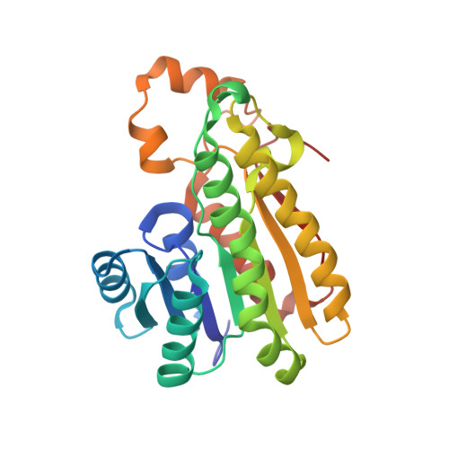
    beta-Ketoacyl-acyl carrier protein reductase (FabG) is a key component in the type II fatty acid synthase system. The structures of Escherichia coli FabG and the FabG[Y151F] mutant in binary complexes with NADP(H) reveal that mechanistically important conformational changes accompany cofactor binding. The active site Ser-Tyr-Lys triad is repositioned into a catalytically competent constellation, and a hydrogen bonded network consisting of ribose hydroxyls, the Ser-Tyr-Lys triad, and four water molecules creates a proton wire to replenish the tyrosine proton donated during catalysis. Also, a disordered loop in FabG forms a substructure in the complex that shapes the entrance to the active site. A key observation is that the nicotinamide portion of the cofactor is disordered in the FabG[Y151F].NADP(H) complex, and Tyr151 appears to be necessary for high-affinity cofactor binding. Biochemical data confirm that FabG[Y151F] is defective in NADPH binding. Finally, structural changes consistent with the observed negative cooperativity of FabG are described.


  • Organizational Affiliation

    Department of Structural Biology, St Jude Children's Research Hospital, Memphis, TN 38105 USA.


Macromolecules
Find similar proteins by:  (by identity cutoff)  |  3D Structure
Entity ID: 1
MoleculeChains Sequence LengthOrganismDetailsImage
3-oxoacyl-[acyl-carrier protein] reductase
A, B
244Escherichia coliMutation(s): 1 
EC: 1.1.1.100
UniProt
Find proteins for P0AEK2 (Escherichia coli (strain K12))
Explore P0AEK2 
Go to UniProtKB:  P0AEK2
Entity Groups  
Sequence Clusters30% Identity50% Identity70% Identity90% Identity95% Identity100% Identity
UniProt GroupP0AEK2
Sequence Annotations
Expand
  • Reference Sequence
Small Molecules
Ligands 1 Unique
IDChains Name / Formula / InChI Key2D Diagram3D Interactions
NDP
Query on NDP

Download Ideal Coordinates CCD File 
C [auth A],
D [auth B]
NADPH DIHYDRO-NICOTINAMIDE-ADENINE-DINUCLEOTIDE PHOSPHATE
C21 H30 N7 O17 P3
ACFIXJIJDZMPPO-NNYOXOHSSA-N
Experimental Data & Validation

Experimental Data

  • Method: X-RAY DIFFRACTION
  • Resolution: 2.50 Å
  • R-Value Free: 0.252 
  • R-Value Work: 0.215 
  • R-Value Observed: 0.219 
  • Space Group: C 2 2 21
Unit Cell:
Length ( Å )Angle ( ˚ )
a = 75.857α = 90
b = 95.857β = 90
c = 131.617γ = 90
Software Package:
Software NamePurpose
PROTEUM PLUSdata collection
SAINTdata reduction
LSCALEdata reduction
AMoREphasing
CNSrefinement
PROTEUM PLUSdata reduction
SAINTdata scaling
LSCALEdata scaling

Structure Validation

View Full Validation Report


Ligand Structure Quality Assessment 


Entry History 

Deposition Data

Revision History  (Full details and data files)

  • Version 1.0: 2004-02-17
    Type: Initial release
  • Version 1.1: 2008-04-29
    Changes: Version format compliance
  • Version 1.2: 2011-07-13
    Changes: Derived calculations, Version format compliance
  • Version 1.3: 2013-11-20
    Changes: Non-polymer description
  • Version 1.4: 2017-10-11
    Changes: Refinement description
  • Version 1.5: 2021-10-27
    Changes: Database references, Derived calculations
  • Version 1.6: 2024-02-21
    Changes: Data collection