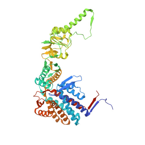
1Q3Q

Crystal structure of the chaperonin from Thermococcus strain KS-1 (two-point mutant complexed with AMP-PNP)


Experimental Data Snapshot

  • Method: X-RAY DIFFRACTION
  • Resolution: 2.30 Å
  • R-Value Free: 0.250 
  • R-Value Work: 0.213 
  • R-Value Observed: 0.213 

Starting Model: experimental
View more details

wwPDB Validation   3D Report Full Report


This is version 1.4 of the entry. See complete history


Literature

Crystal Structures of the Group II Chaperonin from Thermococcus strain KS-1: Steric Hindrance by the Substituted Amino Acid, and Inter-subunit Rearrangement between Two Crystal Forms.

Shomura, Y.Yoshida, T.Iizuka, R.Maruyama, T.Yohda, M.Miki, K.

(2004) J Mol Biol 335: 1265-1278

  • DOI: https://doi.org/10.1016/j.jmb.2003.11.028
  • Primary Citation of Related Structures:  
    1Q2V, 1Q3Q, 1Q3R, 1Q3S

  • PubMed Abstract: 

    The crystal structures of the group II chaperonins consisting of the alpha subunit with amino acid substitutions of G65C and/or I125T from the hyperthermophilic archaeum Thermococcus strain KS-1 were determined. These mutants have been shown to be active in ATP hydrolysis but inactive in protein folding. The structures were shown to be double-ring hexadecamers in an extremely closed form, which was consistent with the crystal structure of native alpha8beta8-chaperonin from Thermoplasma acidophilum. Comparisons of the present structures with the atomic structures of the GroEL14-GroES7-(ADP)7 complex revealed that the deficiency in protein-folding activity with the G65C amino acid substitution is caused by the steric hindrance of the local conformational change in an equatorial domain. We concluded that this mutant chaperonin with G65C substitution is deprived of the smooth conformational change in the refolding-reaction cycle. We obtained a new form of crystal with a distinct space group at a lower concentration of sulfate ion in the presence of nucleotide. The crystal structure obtained at the lower concentration of sulfate ion tilts outward, and has much looser inter-subunit contacts compared with those in the presence of a higher concentration of sulfate ion. Such subunit rotation has never been characterized in group II chaperonins. The crystal structure obtained at the lower concentration of sulfate ion tilts outward, and has much looser inter-subunit contacts compared with those in the presence of a higher concentration of sulfate ion.


  • Organizational Affiliation

    Department of Chemistry, Graduate School of Science, Kyoto University, Sakyo-ku, Kyoto 606-8502, Japan.


Macromolecules
Find similar proteins by:  (by identity cutoff)  |  3D Structure
Entity ID: 1
MoleculeChains Sequence LengthOrganismDetailsImage
Thermosome alpha subunit
A, B, C, D
548Thermococcus sp. JCM 11816Mutation(s): 2 
Gene Names: THSA OR CPKA
EC: 3.6.4.9
UniProt
Find proteins for P61112 (Thermococcus sp. (strain JCM 11816 / KS-1))
Explore P61112 
Go to UniProtKB:  P61112
Entity Groups  
Sequence Clusters30% Identity50% Identity70% Identity90% Identity95% Identity100% Identity
UniProt GroupP61112
Sequence Annotations
Expand
  • Reference Sequence
Small Molecules
Ligands 2 Unique
IDChains Name / Formula / InChI Key2D Diagram3D Interactions
ANP
Query on ANP

Download Ideal Coordinates CCD File 
F [auth A],
H [auth B],
J [auth C],
L [auth D]
PHOSPHOAMINOPHOSPHONIC ACID-ADENYLATE ESTER
C10 H17 N6 O12 P3
PVKSNHVPLWYQGJ-KQYNXXCUSA-N
MG
Query on MG

Download Ideal Coordinates CCD File 
E [auth A],
G [auth B],
I [auth C],
K [auth D]
MAGNESIUM ION
Mg
JLVVSXFLKOJNIY-UHFFFAOYSA-N
Experimental Data & Validation

Experimental Data

  • Method: X-RAY DIFFRACTION
  • Resolution: 2.30 Å
  • R-Value Free: 0.250 
  • R-Value Work: 0.213 
  • R-Value Observed: 0.213 
  • Space Group: P 4 21 2
Unit Cell:
Length ( Å )Angle ( ˚ )
a = 210.829α = 90
b = 210.829β = 90
c = 156.421γ = 90
Software Package:
Software NamePurpose
CNSrefinement
DENZOdata reduction
SCALEPACKdata scaling
AMoREphasing

Structure Validation

View Full Validation Report


Entry History 

Deposition Data

Revision History  (Full details and data files)

  • Version 1.0: 2004-01-27
    Type: Initial release
  • Version 1.1: 2008-04-29
    Changes: Version format compliance
  • Version 1.2: 2011-07-13
    Changes: Derived calculations, Source and taxonomy, Version format compliance
  • Version 1.3: 2021-10-27
    Changes: Database references, Derived calculations
  • Version 1.4: 2023-08-16
    Changes: Data collection, Refinement description