1Q35 | pdb_00001q35

Crystal Structure of Pasteurella haemolytica Apo Ferric ion-Binding Protein A


Experimental Data Snapshot

  • Method: X-RAY DIFFRACTION
  • Resolution: 1.20 Å
  • R-Value Free: 
    0.195 (Depositor), 0.207 (DCC) 
  • R-Value Work: 
    0.171 (Depositor), 0.181 (DCC) 
  • R-Value Observed: 
    0.172 (Depositor) 

wwPDB Validation 3D Report Full Report

Validation slider image for 1Q35

This is version 1.3 of the entry. See complete history

Literature

Crystal structure of Pasteurella haemolytica ferric ion-binding protein A reveals a novel class of bacterial iron-binding proteins

Shouldice, S.R.Dougan, D.R.Williams, P.A.Skene, R.J.Snell, G.Scheibe, D.Kirby, S.Hosfield, D.J.McRee, D.E.Schryvers, A.B.Tari, L.W.

(2003) J Biological Chem 278: 41093-41098

  • DOI: https://doi.org/10.1074/jbc.M306821200
  • Primary Citation Related Structures: 
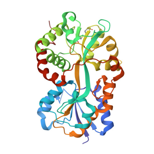
    1Q35

  • PubMed Abstract: 

    Pasteurellosis caused by the Gram-negative pathogen Pasteurella haemolytica is a serious disease leading to death in cattle. To scavenge growth-limiting iron from the host, the pathogen utilizes the periplasmic ferric ion-binding protein A (PhFbpA) as a component of an ATP-binding cassette transport pathway. We report the 1.2-A structure of the iron-free (apo) form of PhFbpA, which is a member of the transferrin structural superfamily. The protein structure adopts a closed conformation, allowing us to reliably assign putative iron-coordinating residues. Based on our analysis, PhFbpA utilizes a unique constellation of binding site residues and anions to octahedrally coordinate an iron atom. A surprising finding in the structure is the presence of two formate anions on opposite sides of the iron-binding pocket. The formate ions tether the N- and C-terminal domains of the protein and stabilize the closed structure, also providing clues as to probable candidates for synergistic anions in the iron-loaded state. PhFbpA represents a new class of bacterial iron-binding proteins.


  • Organizational Affiliation
    • Department of Microbiology and Infectious Diseases, University of Calgary, Calgary, Alberta T2N 4N1, Canada.

Macromolecule Content 

  • Total Structure Weight: 36.05 kDa 
  • Atom Count: 3,143 
  • Modeled Residue Count: 317 
  • Deposited Residue Count: 320 
  • Unique protein chains: 1

Macromolecules

Find similar proteins by:|  3D Structure
Entity ID: 1
MoleculeChains  Sequence LengthOrganismDetailsImage
iron binding protein FbpA320Mannheimia haemolyticaMutation(s): 0 
Gene Names: fbpA
UniProt
Find proteins for Q9Z4N6 (Mannheimia haemolytica)
Explore Q9Z4N6 
Go to UniProtKB:  Q9Z4N6
Entity Groups
Sequence Clusters30% Identity50% Identity70% Identity90% Identity95% Identity100% Identity
UniProt GroupQ9Z4N6
Sequence Annotations
Expand
Reference Sequence

Experimental Data & Validation

Experimental Data

  • Method: X-RAY DIFFRACTION
  • Resolution: 1.20 Å
  • R-Value Free:  0.195 (Depositor), 0.207 (DCC) 
  • R-Value Work:  0.171 (Depositor), 0.181 (DCC) 
  • R-Value Observed: 0.172 (Depositor) 
Space Group: C 2 2 21
Unit Cell:
Length ( Å )Angle ( ˚ )
a = 97.159α = 90
b = 189.327β = 90
c = 45.765γ = 90
Software Package:
Software NamePurpose
REFMACrefinement
HKL-2000data reduction
CCP4data scaling
MOLREPphasing

Structure Validation

View Full Validation Report


Entry History 

Deposition Data

Revision History  (Full details and data files)

  • Version 1.0: 2003-11-11
    Type: Initial release
  • Version 1.1: 2008-04-29
    Changes: Version format compliance
  • Version 1.2: 2011-07-13
    Changes: Version format compliance
  • Version 1.3: 2024-11-13
    Changes: Data collection, Database references, Derived calculations, Structure summary