1Q23 | pdb_00001q23

Crystal structure of Chloramphenicol acetyltransferase I complexed with Fusidic acid at 2.18 A resolution


Experimental Data Snapshot

  • Method: X-RAY DIFFRACTION
  • Resolution: 2.18 Å
  • R-Value Free: 
    0.263 (Depositor), 0.265 (DCC) 
  • R-Value Work: 
    0.192 (Depositor), 0.200 (DCC) 
  • R-Value Observed: 
    0.195 (Depositor) 

Starting Model: experimental
View more details

wwPDB Validation 3D Report Full Report

Validation slider image for 1Q23

Ligand Structure Quality Assessment 


This is version 1.3 of the entry. See complete history

Literature

Crystal structure of Chloramphenicol acetyltransferase I in the apoenzyme form and complexed with fusidic acid at 2.18 A resolution

Roidis, A.Kokkinidis, M.

To be published.

Macromolecule Content 

  • Total Structure Weight: 314.55 kDa 
  • Atom Count: 22,280 
  • Modeled Residue Count: 2,563 
  • Deposited Residue Count: 2,628 
  • Unique protein chains: 1

Macromolecules

Find similar proteins by:|  3D Structure
Entity ID: 1
MoleculeChains  Sequence LengthOrganismDetailsImage
Chloramphenicol acetyltransferase
A, B, C, D, E
A, B, C, D, E, F, G, H, I, J, K, L
219Escherichia coliMutation(s): 0 
Gene Names: CAT OR HCM1.206
EC: 2.3.1.28
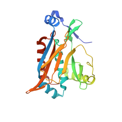
UniProt
Find proteins for P62577 (Escherichia coli)
Explore P62577 
Go to UniProtKB:  P62577
Entity Groups
Sequence Clusters30% Identity50% Identity70% Identity90% Identity95% Identity100% Identity
UniProt GroupP62577
Sequence Annotations
Expand
Reference Sequence

Small Molecules

Ligands 2 Unique
IDChains Name / Formula / InChI Key2D Diagram3D Interactions
FUA

Query on FUA


Download:Ideal Coordinates CCD File
N [auth A]
O [auth B]
P [auth C]
Q [auth D]
R [auth E]
N [auth A],
O [auth B],
P [auth C],
Q [auth D],
R [auth E],
S [auth F],
T [auth G],
U [auth H],
V [auth I],
W [auth J],
X [auth K],
Y [auth L]
FUSIDIC ACID
C31 H48 O6
IECPWNUMDGFDKC-MZJAQBGESA-N
CA

Query on CA


Download:Ideal Coordinates CCD File
M [auth A]CALCIUM ION
Ca
BHPQYMZQTOCNFJ-UHFFFAOYSA-N

Experimental Data & Validation

Experimental Data

  • Method: X-RAY DIFFRACTION
  • Resolution: 2.18 Å
  • R-Value Free:  0.263 (Depositor), 0.265 (DCC) 
  • R-Value Work:  0.192 (Depositor), 0.200 (DCC) 
  • R-Value Observed: 0.195 (Depositor) 
Space Group: P 1 21 1
Unit Cell:
Length ( Å )Angle ( ˚ )
a = 115.354α = 90
b = 129.198β = 108.3
c = 118.073γ = 90
Software Package:
Software NamePurpose
REFMACrefinement
MOSFLMdata reduction
CCP4data scaling
AMoREphasing

Structure Validation

View Full Validation Report


Ligand Structure Quality Assessment 


Entry History 

Deposition Data

Revision History  (Full details and data files)

  • Version 1.0: 2004-08-03
    Type: Initial release
  • Version 1.1: 2008-04-29
    Changes: Version format compliance
  • Version 1.2: 2011-07-13
    Changes: Version format compliance
  • Version 1.3: 2023-08-16
    Changes: Data collection, Database references, Derived calculations, Refinement description