Crystal structure of a novel human peroxidase enzyme at 2.0 A resolution.
Choi, H.J., Kang, S.W., Yang, C.H., Rhee, S.G., Ryu, S.E.(1998) Nat Struct Biol 5: 400-406
- PubMed: 9587003 Search on PubMed
- DOI: https://doi.org/10.1038/nsb0598-400
- Primary Citation Related Structures:
1PRX - PubMed Abstract:
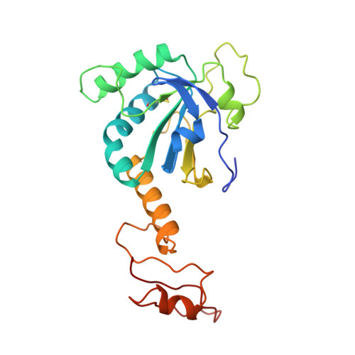
Hydrogen peroxide (H2O2) has been implicated recently as an intracellular messenger that affects cellular processes including protein phosphorylation, transcription and apoptosis. A set of novel peroxidases, named peroxiredoxins (Prx), regulate the intracellular concentration of H2O2 by reducing it in the presence of an appropriate electron donor. The crystal structure of a human Prx enzyme, hORF6, reveals that the protein contains two discrete domains and forms a dimer. The N-terminal domain has a thioredoxin fold and the C-terminal domain is used for dimerization. The active site cysteine (Cys 47), which exists as cysteine-sulfenic acid in the crystal, is located at the bottom of a relatively narrow pocket. The positively charged environment surrounding Cys 47 accounts for the peroxidase activity of the enzyme, which contains no redox cofactors.
- Division of Protein Engineering, Korea Research Institute of Bioscience and Biotechnology, KIST, Yusong, Taejon, South Korea.
Organizational Affiliation: