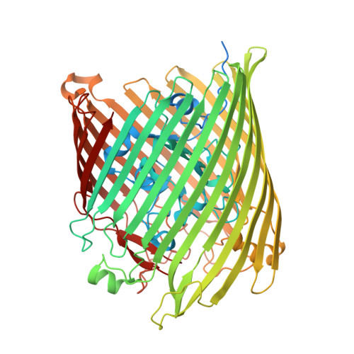
Structural evidence for iron-free citrate and ferric citrate binding to the TonB-dependent outer membrane transporter FecA
Yue, W.W., Grizot, S., Buchanan, S.K.(2003) J Mol Biology 332: 353-368
- PubMed: 12948487
- DOI: https://doi.org/10.1016/s0022-2836(03)00855-6
- Primary Citation of Related Structures:
1PNZ, 1PO0, 1PO3 - PubMed Abstract:
Escherichia coli possesses a TonB-dependent transport system, which exploits the iron-binding capacity of citrate and its natural abundance. Here, we describe three structures of the outer membrane ferric citrate transporter FecA: unliganded and complexed with iron-free or diferric dicitrate. We show the structural mechanism for discrimination between the iron-free and ferric siderophore: the binding of diferric dicitrate, but not iron-free dicitrate alone, causes major conformational rearrangements in the transporter. The structure of FecA bound with iron-free dicitrate represents the first structure of a TonB-dependent transporter bound with an iron-free siderophore. Binding of diferric dicitrate to FecA results in changes in the orientation of the two citrate ions relative to each other and in their interactions with FecA, compared to the binding of iron-free dicitrate. The changes in ligand binding are accompanied by conformational changes in three areas of FecA: two extracellular loops, one plug domain loop and the periplasmic TonB-box motif. The positional and conformational changes in the siderophore and transporter initiate two independent events: ferric citrate transport into the periplasm and transcription induction of the fecABCDE transport genes. From these data, we propose a two-step ligand recognition event: FecA binds iron-free dicitrate in the non-productive state or first step, followed by siderophore displacement to form the transport-competent, diferric dicitrate-bound state in the second step.
- National Institute of Diabetes and Digestive and Kidney Diseases, National Institutes of Health, Bethesda, MD 20892, USA.
Organizational Affiliation:


