1PGU | pdb_00001pgu

YEAST ACTIN INTERACTING PROTEIN 1 (AIP1), Se-Met PROTEIN, MONOCLINIC CRYSTAL FORM


Experimental Data Snapshot

  • Method: X-RAY DIFFRACTION
  • Resolution: 2.30 Å
  • R-Value Free: 
    0.257 (Depositor) 
  • R-Value Work: 
    0.202 (Depositor) 
  • R-Value Observed: 
    0.202 (Depositor) 

wwPDB Validation 3D Report Full Report

Validation slider image for 1PGU

This is version 1.4 of the entry. See complete history

Literature

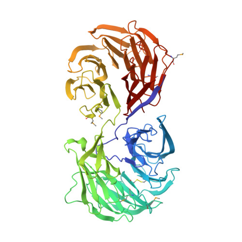
The structure of Aip1p, a WD repeat protein that regulates Cofilin-mediated actin depolymerization.

Voegtli, W.C.Madrona, A.Y.Wilson, D.K.

(2003) J Biological Chem 278: 34373-34379

  • DOI: https://doi.org/10.1074/jbc.M302773200
  • Primary Citation Related Structures: 
    1PGU, 1PI6

  • PubMed Abstract: 

    Actin-interacting protein 1 (Aip1p) is a 67-kDa WD repeat protein known to regulate the depolymerization of actin filaments by cofilin and is conserved in organisms ranging from yeast to mammals. The crystal structure of Aip1p from Saccharomyces cerevisiae was determined to a 2.3-A resolution and a final crystallographic R-factor of 0.204. The structure reveals that the overall fold is formed by two connected seven-bladed beta-propellers and has important implications for the structure of Aip1 from other organisms and WD repeat-containing proteins in general. These results were unexpected because a maximum of 10 WD repeats had been reported in the literature for this protein using sequence data. The surfaces of the beta-propellers formed by the D-A and B-C loops are positioned adjacent to one another, giving Aip1p a shape that resembles an open "clamshell." The mapping of conserved residues to the structure of Aip1p reveals dense patches of conserved residues on the surface of one beta-propeller and at the interface of the two beta-propellers. These two patches of conserved residues suggest a potential binding site for F-actin on Aip1p and that the orientation of the beta-propellers with respect to one another plays a role in binding an actin-cofilin complex. In addition, the conserved interface between the domains is mediated by a number of interactions that appear to impart rigidity between the two domains of Aip1p and may make a large substrate-induced conformational change difficult.


  • Organizational Affiliation
    • Section of Molecular and Cellular Biology, University of California, Davis, California 95616, USA.

Macromolecule Content 

  • Total Structure Weight: 135.52 kDa 
  • Atom Count: 9,844 
  • Modeled Residue Count: 1,214 
  • Deposited Residue Count: 1,230 
  • Unique protein chains: 1

Macromolecules

Find similar proteins by:|  3D Structure
Entity ID: 1
MoleculeChains  Sequence LengthOrganismDetailsImage
Actin interacting protein 1
A, B
615Saccharomyces cerevisiaeMutation(s): 7 
Gene Names: AIP1 OR YMR092C OR YM9582.17C
UniProt
Find proteins for P46680 (Saccharomyces cerevisiae (strain ATCC 204508 / S288c))
Explore P46680 
Go to UniProtKB:  P46680
Entity Groups
Sequence Clusters30% Identity50% Identity70% Identity90% Identity95% Identity100% Identity
UniProt GroupP46680
Sequence Annotations
Expand
Reference Sequence

Small Molecules

Modified Residues  1 Unique
IDChains TypeFormula2D DiagramParent
MSE
Query on MSE
A, B
L-PEPTIDE LINKINGC5 H11 N O2 SeMET

Experimental Data & Validation

Experimental Data

  • Method: X-RAY DIFFRACTION
  • Resolution: 2.30 Å
  • R-Value Free:  0.257 (Depositor) 
  • R-Value Work:  0.202 (Depositor) 
  • R-Value Observed: 0.202 (Depositor) 
Space Group: P 1 21 1
Unit Cell:
Length ( Å )Angle ( ˚ )
a = 60.8α = 90
b = 154.53β = 90.65
c = 69γ = 90
Software Package:
Software NamePurpose
CNSrefinement
DENZOdata reduction
SCALEPACKdata scaling
SOLVEphasing
CNSphasing

Structure Validation

View Full Validation Report


Entry History 

Deposition Data

Revision History  (Full details and data files)

  • Version 1.0: 2003-07-15
    Type: Initial release
  • Version 1.1: 2008-04-29
    Changes: Version format compliance
  • Version 1.2: 2011-07-13
    Changes: Version format compliance
  • Version 1.3: 2021-10-27
    Changes: Database references, Derived calculations
  • Version 1.4: 2024-10-30
    Changes: Data collection, Structure summary